Figure 3. Genetic findings. A: Sequence electropherogram displaying the homozygous progressive rod-cone degeneration mutation c.52C>T p.R18X. B: Sequence electropherogram displaying the heterozygous RBP3 mutation c.1694T>C p.V565A. Beneath the electropherogram, the
wild-type and mutant nucleotide-encoded amino acid sequences are shown.
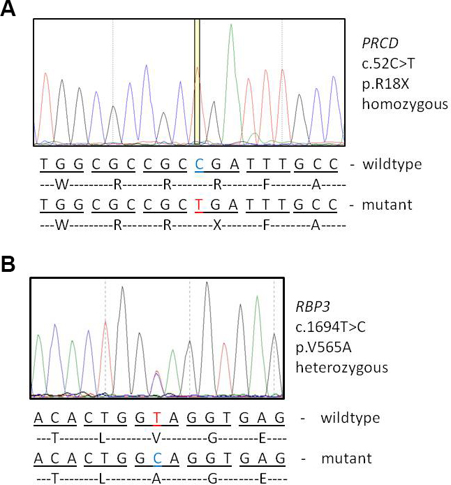
 Figure 3 of
Pach, Mol Vis 2013; 19:1350-1355.
Figure 3 of
Pach, Mol Vis 2013; 19:1350-1355.