Figure 2. Clinical findings of the three siblings carrying a homozygous mutation in the progressive rod-cone degeneration (PRCD) gene. A–C: Perimetry shows visual field constriction in every patient with residual peripheral islands in the two female patients (target
color coding: green: I4e, red: III4e, blue: V4e). D–F: Fundus photography shows bone-spicule-like pigmentation, attenuated arterioles and bull´s eye maculopathy (BEM) due to a
patchy RPE atrophy. G–I: FAF shows sharply demarcated, smaller and larger, partly confluent areas of reduced autofluorescence. J–L: Optical coherence tomography (OCT) imaging shows a decrease in retinal thickness with shortening of the photoreceptor outer
segments, reduction in the outer nuclear layer, and RPE atrophy. (Findings are shown on the left for patient IV:3, in the
middle for patient IV:4, and on the right for patient IV:5.)
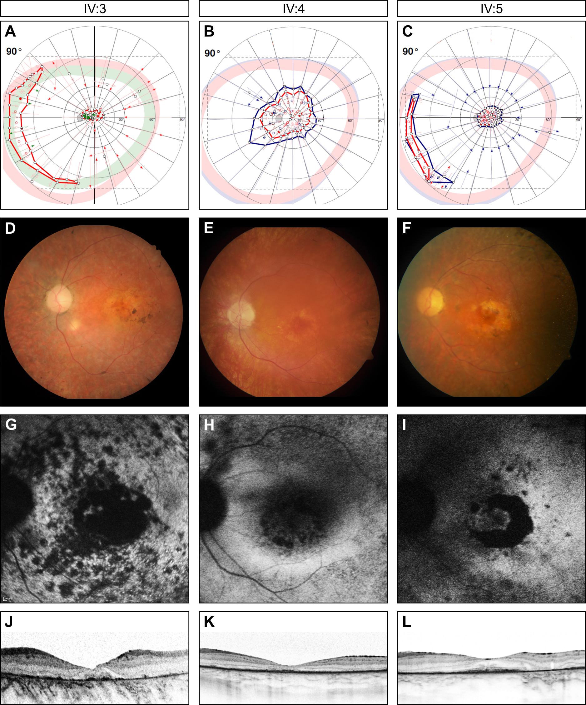
 Figure 2 of
Pach, Mol Vis 2013; 19:1350-1355.
Figure 2 of
Pach, Mol Vis 2013; 19:1350-1355.