Figure 5. Time course of H2O2-induced JNK phosphorylation and NFκB. Porcine trabecular meshwork (PTM) cells were treated with medium alone (control) or
with 600 μM H2O2. Cytosol or nucleus fraction was extracted from TM cells. Cytoplasmic proteins were separated by sodium dodecyl sulfate PAGE,
transferred onto a polyvinylidene fluoride transfer membrane, and probed with antibody to p-JNK, JNK (A), and NFκB (B). Levels protein bands were quantified, and the expression ratios of phosphorylated protein to total protein for JNK and
nucleus to cytosol for NFκB were calculated (n=3). *p<0.05 compared with medium alone. Bars indicate the standard deviations.
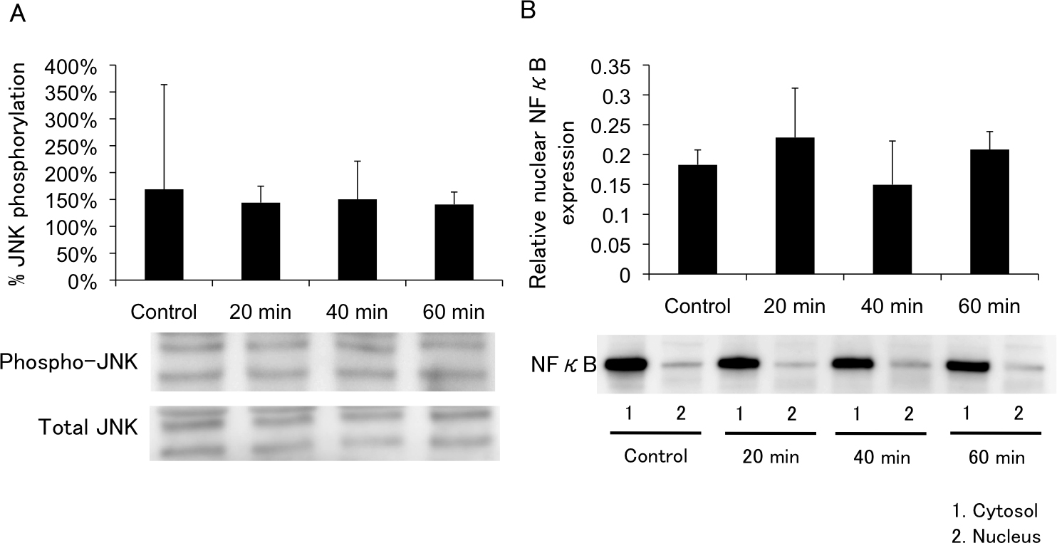
 Figure 5 of
Awai-Kasaoka, Mol Vis 2013; 19:1332-1340.
Figure 5 of
Awai-Kasaoka, Mol Vis 2013; 19:1332-1340.