Figure 4. Time course of H2O2-induced Akt (Ser473), ERK1/2, p38 phosphorylation. Porcine trabecular meshwork (PTM) cells (1x10⁵) in 12-well plates were treated with medium alone (control) or 600 μM H2O2 for various amounts of time. Time course H2O2-induced Akt (Ser473), ERK1/2, p38 phosphorylation were indicated panels A, B, and C, respectively. Levels of protein bands were quantified, and the ratios of phosphorylated protein to total protein were calculated
(n=3). *p<0.05 compared with medium alone. Bars indicate the standard deviations.
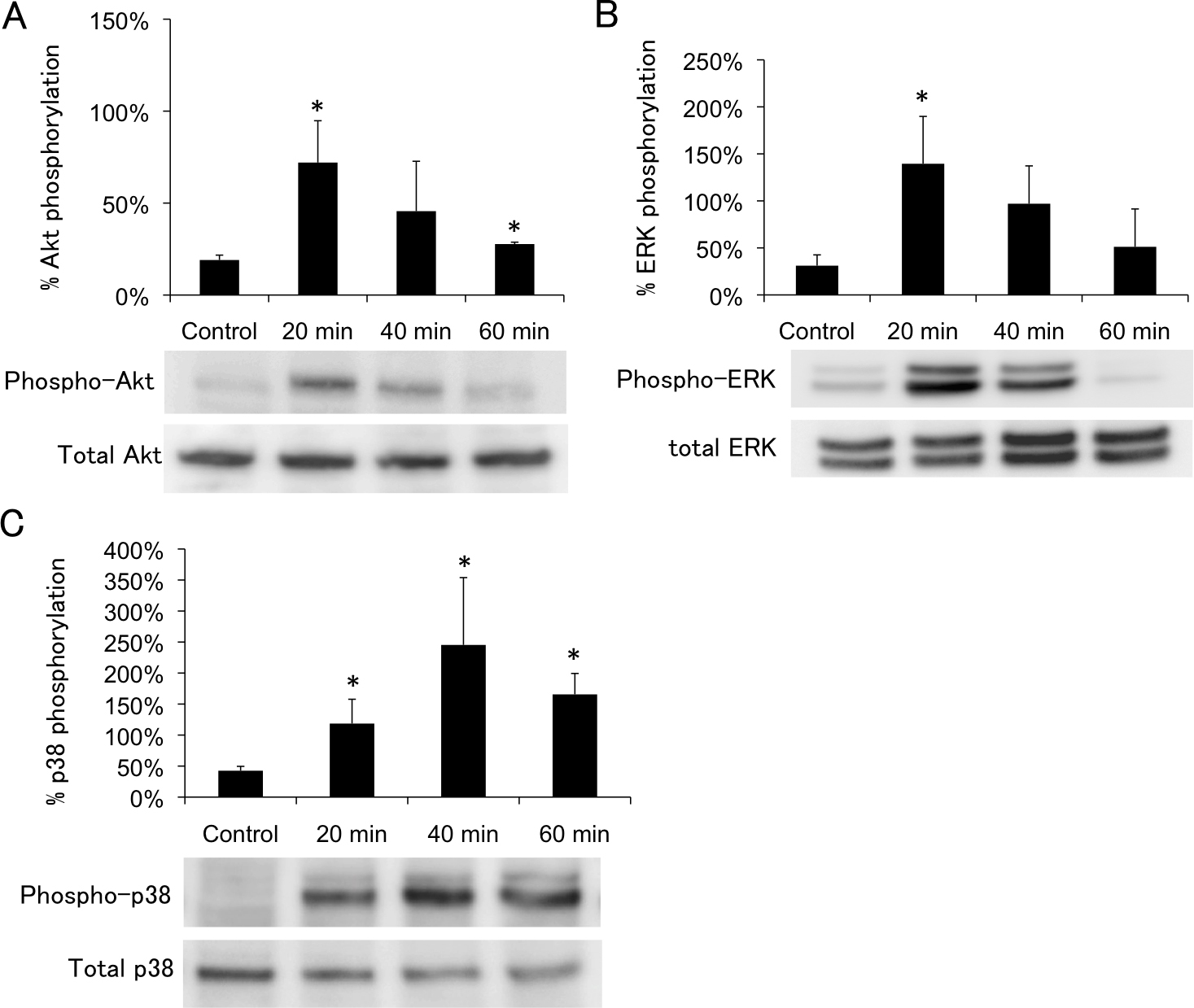
 Figure 4 of
Awai-Kasaoka, Mol Vis 2013; 19:1332-1340.
Figure 4 of
Awai-Kasaoka, Mol Vis 2013; 19:1332-1340.