Figure 2. Immunocytochemical staining of H2O2-treated porcine trabecular meshwork (PTM) cells. PTM cells treated with 600 or 800 μM H2O2 for 6 h were fixed, permeabilized, and stained with anticleaved caspase-3 antibody (green; upper panels). F-actin was stained
with phalloidin-TRITC (red; lower panels). Cell nuclei were counterstained with DAPI (blue). Scale bar represents 50 μm.
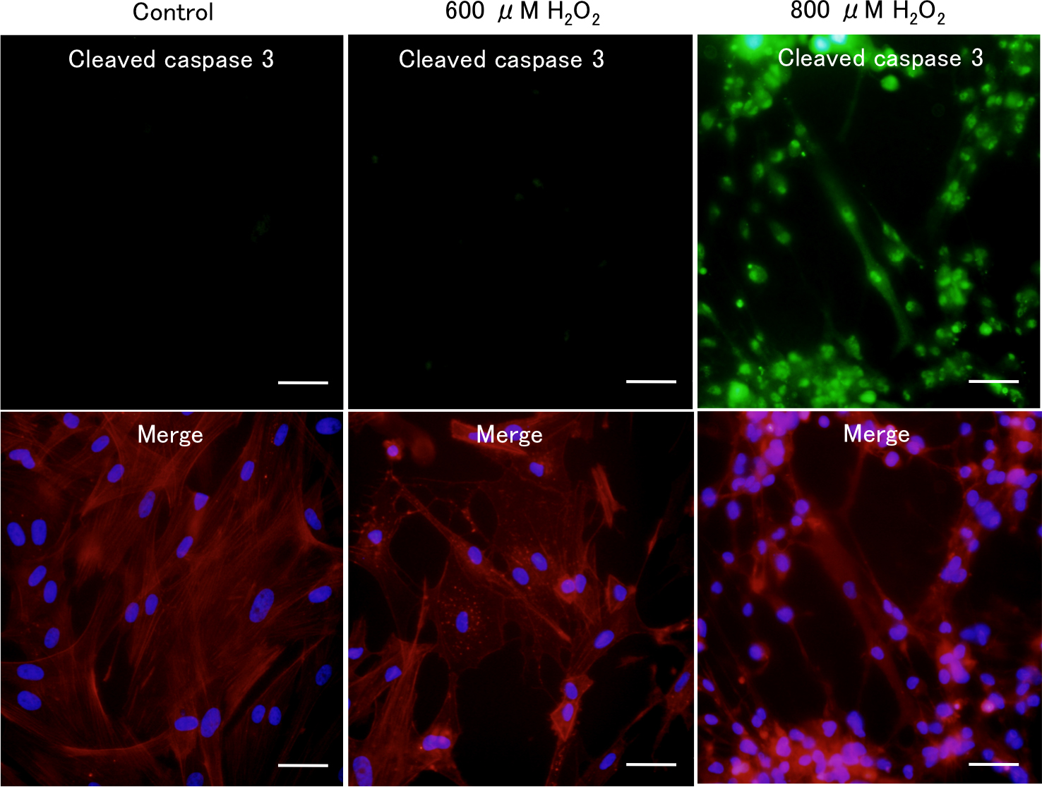
 Figure 2 of
Awai-Kasaoka, Mol Vis 2013; 19:1332-1340.
Figure 2 of
Awai-Kasaoka, Mol Vis 2013; 19:1332-1340.