Figure 1. Cellular morphological changes in porcine trabecular meshwork (PTM) cells treated with medium alone, or with 600 μM or 800
μM H2O2. PTM (1x10⁵) cells were seeded into 12-well plates. Three days later, the medium was changed to serum-free medium overnight, and then
PTM cells were treated as followed for various amounts of time: A: medium alone, B: 600 μM H2O2C: 800 μM H2O2. The unfixed cells were stained with propidium iodide to detect cell death (red; middle panel). Nuclei of total cells were
stained with Hoechst 33342 (blue; lower panel). The time course for mean percentages of dead cells (propidium iodide-positive
cells) to total number of cells (Hoechst 33342-positive cells) is shown (n=3; D). Thus, 800 μM H2O2 increased the number of dead PTM cells for 8 h after treatment, but PTM cells treated with 600 μM H2O2 recovered from the morphologic changes without significant cell death. Scale bar represents 50 μm.
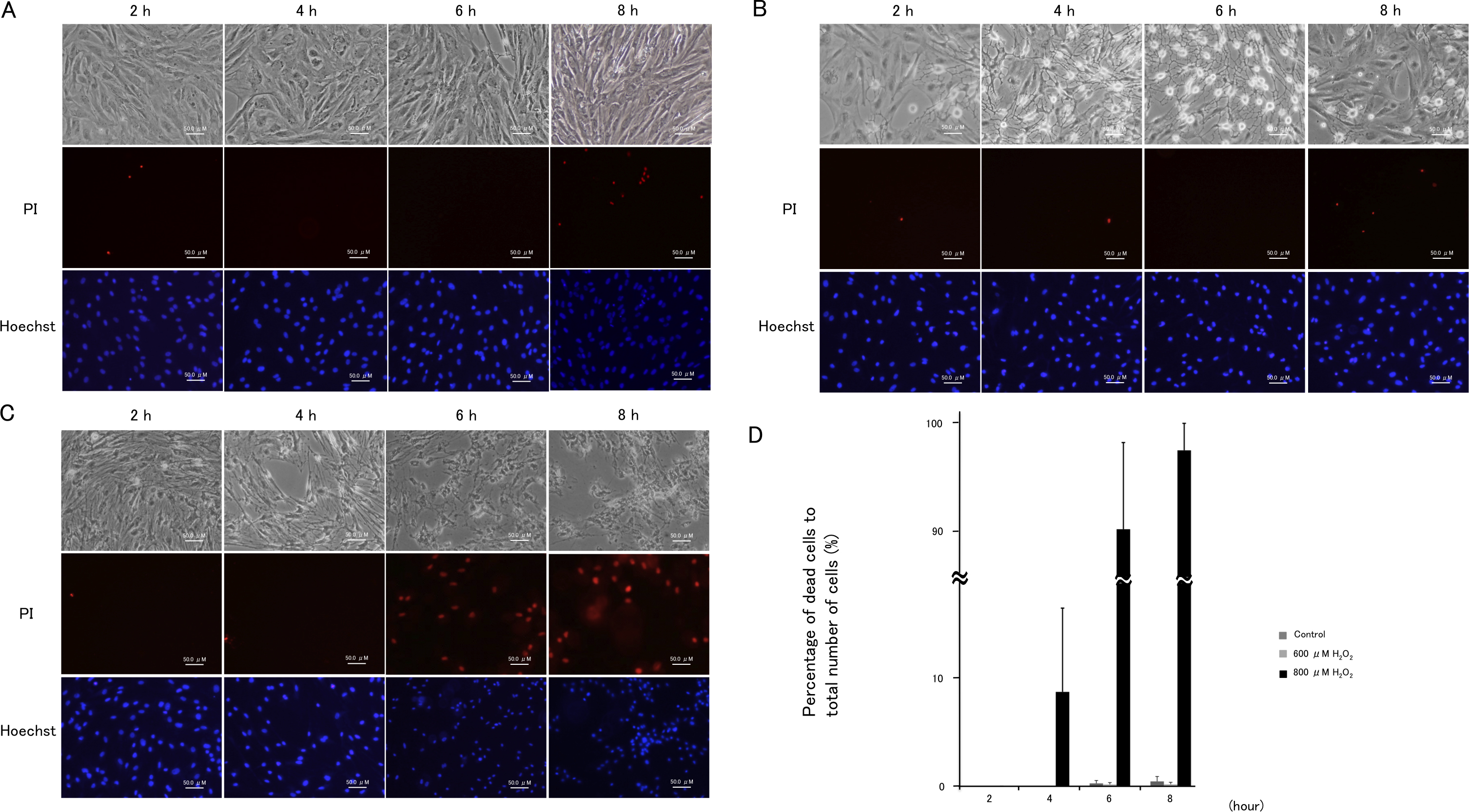
 Figure 1 of
Awai-Kasaoka, Mol Vis 2013; 19:1332-1340.
Figure 1 of
Awai-Kasaoka, Mol Vis 2013; 19:1332-1340.