Figure 3. Histology and immunohistochemistry. A: The Cross section of the eyeball was stained for connective tissue. B: The Cross section of the eyeball was stained for collagen I. C: The cross section through the conjuntival fornix was stained for connective tissue. D: The cross section through the conjunctival fornix was stained for collagen I. Collagen I is absent from the retina and the
conjunctival epithelium (e) but present in the small region of Tenon’s space (T), the choroid, and the sclera.
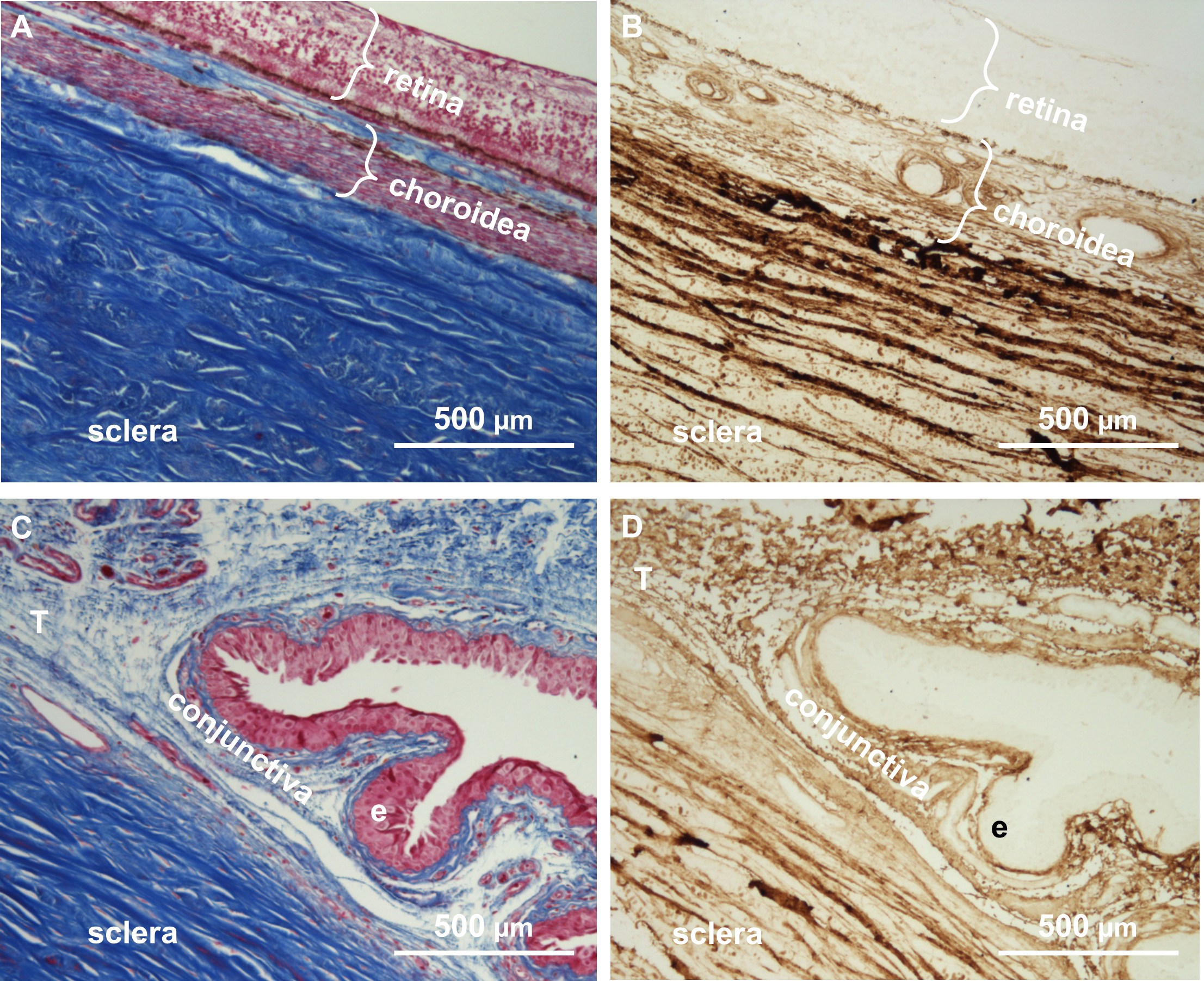
 Figure 3 of
Löbler, Mol Vis 2013; 19:1321-1331.
Figure 3 of
Löbler, Mol Vis 2013; 19:1321-1331.