Figure 4. The tdTomato-expressing retinal bipolar cells in the Pcp2-cre mouse line are co-labeled with antibodies specific to rod bipolar
cells, type 2 and 6 cone bipolar cells. A–C: In retinal vertical sections, the tdTomato-expressing retina (A) was immunostained for PKCα (B). The overlay of A and B is shown in C. The double-positive bipolar cells were marked with stars in the somata and with arrowheads pointing at the axon terminals.
D–F: In the retinal whole mount with the focal plane in the INL, colabeling with tdTomato and PKCα. PKCα-negative tdTomato-expressing
cells are marked with arrows. G–I: The tdTomato-expressing retina was immunostained for Syt2. The double-positive bipolar cells with axon terminals stratified
at the distal portion of the IPL (type 2 bipolar cells) are marked with white stars in the somata and with white arrowheads
pointing at the axon terminals. The double-positive bipolar cells with axon terminals stratified in the proximal portion of
the IPL (type 6 bipolar cells) are marked with yellow arrows at the somata and yellow arrowheads at the axon terminals. A
tdTomato-expressing bipolar cell with their axon terminals stratified slightly distal to Syt2-positive cells is marked with
a blue arrowhead (I). The tdTomato-expressing retinal bipolar cells were not found to be labeled by PKARIIβ (J–L), HCN4 (M–O), and calsenilin (P–R). Scale bars represent 50 µm. ONL, outer nuclear layer; INL, inner nuclear layer; IPL, inner plexiform layer; GCL, ganglion
cell layer.
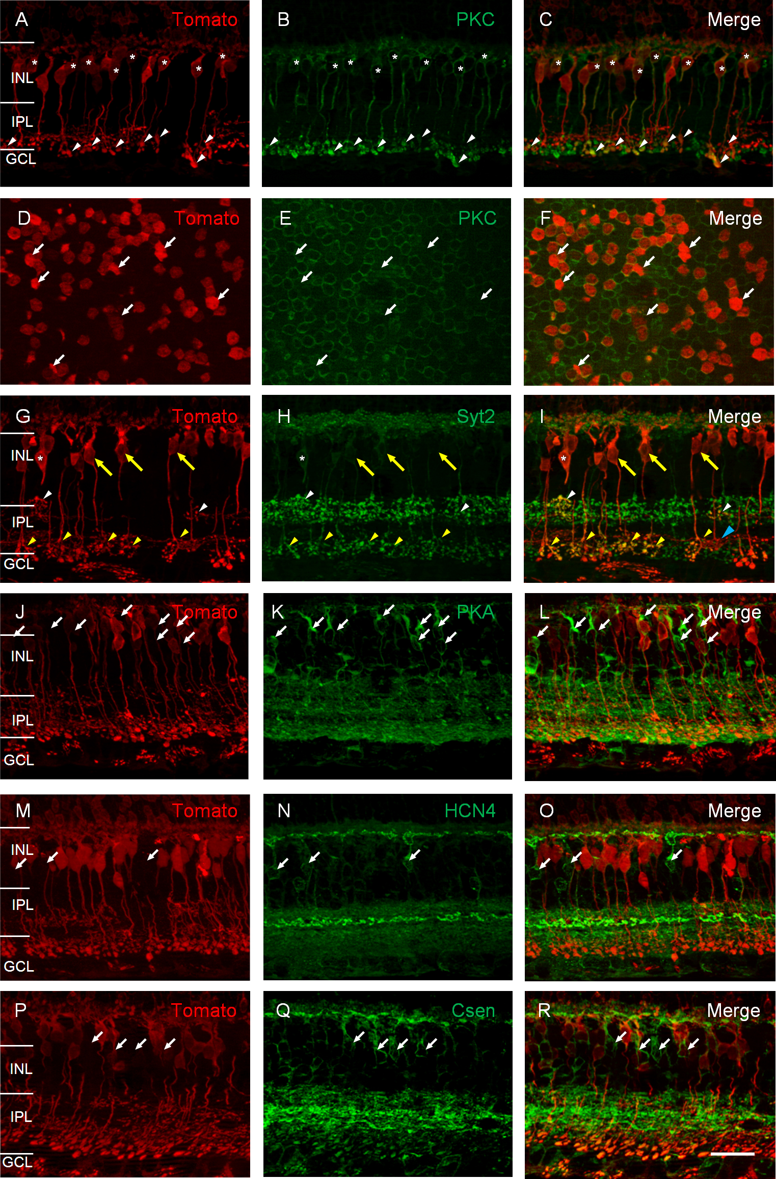
 Figure 4 of
Lu, Mol Vis 2013; 19:1310-1320.
Figure 4 of
Lu, Mol Vis 2013; 19:1310-1320.