Figure 3. tdTomato-expressing bipolar cells in the Pcp2-cre transgenic mouse line. B: Representative bipolar cells in high magnification viewed in retinal vertical sections. The inner plexiform layer (IPL)
was divided into five sublaminae. White arrowheads point to the axon terminals of rod bipolar cells. Yellow arrowheads point
to the axon terminals of bipolar cells stratified in the distal portion of the IPL. Blue arrowheads point to the axon terminals
of bipolar cells stratified in the proximal portion of the IPL. C: tdTomato-expressing cells viewed in a retinal whole mount with the focal plane at the INL. D: Whole-mount view of the axon terminals of the tdTomato-expressing bipolar cells with the focal plane at the proximal portion
of the IPL to the ganglion cell layer. The arrowhead points to a weak tdTomato-expressing ganglion cell. The images in C and D were taken in the same field. Scale bars represent 25 µm in A, C, and D and 10 µm in B. ONL, outer nuclear layer; INL, inner nuclear layer; GCL, ganglion cell layer.
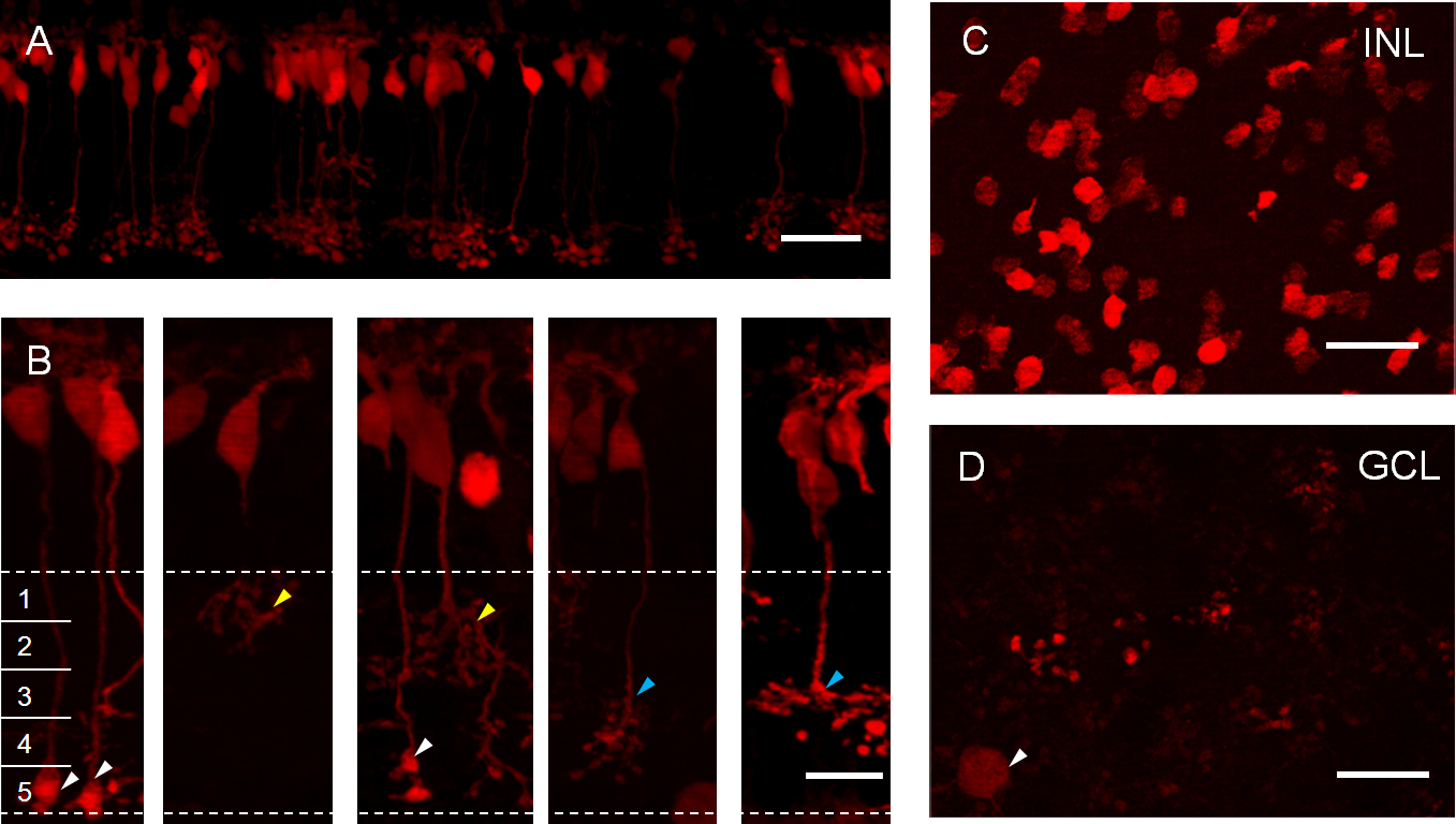
 Figure 3 of
Lu, Mol Vis 2013; 19:1310-1320.
Figure 3 of
Lu, Mol Vis 2013; 19:1310-1320.