Figure 2. Disease-associated mutations in CNGB3 increase cytotoxicity. A: Western blot demonstrating expression of FLAG-tagged wild-type (WT) and mutant cone CNG channel subunits in 661W cells following
transfection with indicated plasmids (above). Approximate locations of molecular weight markers (in kilodaltons) are indicated
to the right of the immunoblot. Cell lysates were also probed with beta-actin antibody (below). The molecular weight of beta
actin was ~42 kDa. B: The bar graph demonstrates increased cytotoxicity (measured as LDH release from dying cells) for cells expressing CNGA3
plus wild-type (WT) or mutant CNGB3 exposed to various concentrations of CPT-cGMP for 24 h (n=46 to 48). Cytotoxicity was
normalized to that of control cells transfected with vector (pOPRSVI) alone, incubated in the absence of CPT-cGMP; *, significant
difference between groups indicated by bracket (p<0.05); +, significant difference between F525N groups with or without 0.1
μM CPT-cGMP treatment (p<0.01).
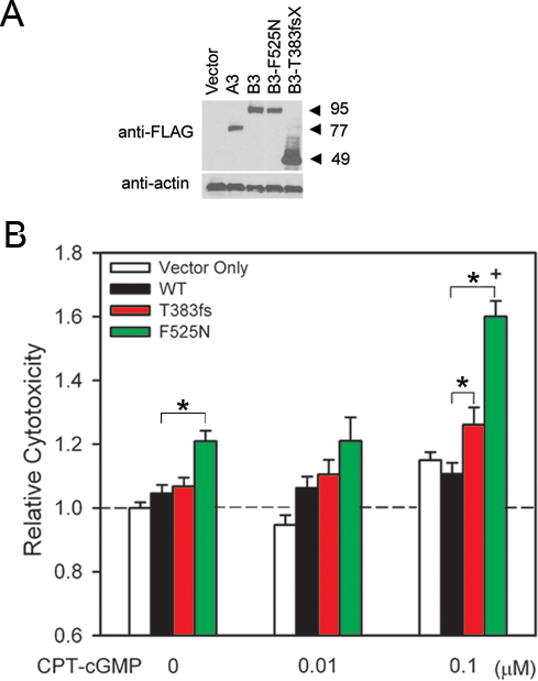
 Figure 2 of
Liu, Mol Vis 2013; 19:1268-1281.
Figure 2 of
Liu, Mol Vis 2013; 19:1268-1281.