Figure 7. Scanning electron microscopy. At day 1, some corneal endothelial cells (CECs) are still attached to Descemet’s membrane (DM)
in both groups, although most of the cells were ablated in both groups. Most of the remnant cells have detached between day
3 and day 14 in both groups. At day 21, denuded DM with only scant distribution of remnant CECs was noted in both groups.
Scale bar=20 μm. *taken from the opposite eye that did not undergo cryoinjury.
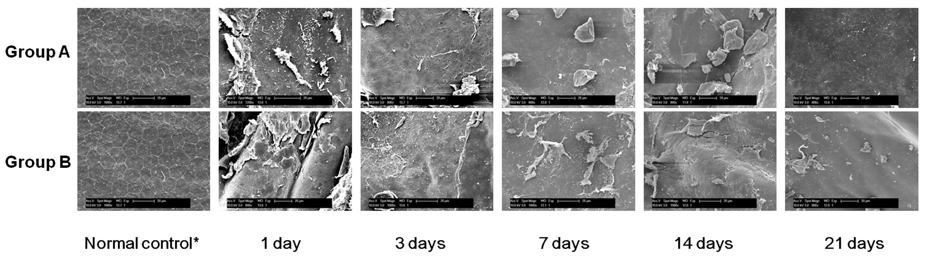
 Figure 7 of
Han, Mol Vis 2013; 19:1222-1230.
Figure 7 of
Han, Mol Vis 2013; 19:1222-1230.