Figure 5. Hematoxylin and eosin staining. Ablation of endothelial cells was observed at all time points in both groups. At 1 day after
cryoinjury, more severe infiltration of inflammatory cells throughout all layers of the cornea in group B compared to that
in group A was observed. Although Descemet’s membrane (DM) was mostly preserved, detachment of DM was detected in some areas
(black arrow). Scale bar=50 μm. *taken from the opposite eye that did not undergo cryoinjury.
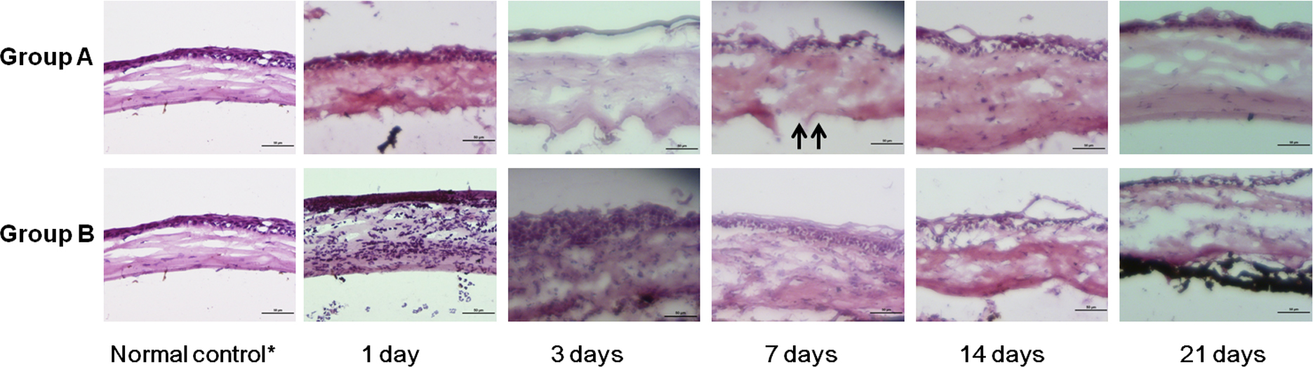
 Figure 5 of
Han, Mol Vis 2013; 19:1222-1230.
Figure 5 of
Han, Mol Vis 2013; 19:1222-1230.