Figure 4. Live/dead cell assay using alizarin red S and Trypan Blue staining of the corneal endothelial cells. Successful ablation of
the corneal endothelial cells was observed in samples from day 1, and no evidence of cell regrowth was detected during the
study period. Descemet’s membrane was preserved over most of the area. Scale bar=50 μm. *taken from the opposite eye that
did not undergo cryoinjury.
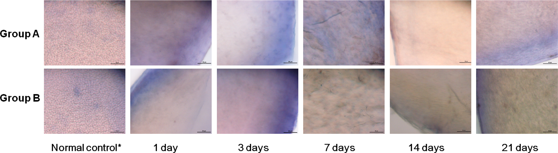
 Figure 4 of
Han, Mol Vis 2013; 19:1222-1230.
Figure 4 of
Han, Mol Vis 2013; 19:1222-1230.