Figure 2. Anterior segment optical coherence tomography. Significant corneal edema developed in groups A and B, and the increased corneal
thickness was maintained during the study period. Although the anterior segment appears normal except the corneal edema and
mild cataract in group A, severe anterior chamber inflammation and iridocorneal adhesion are remarkable in group B.
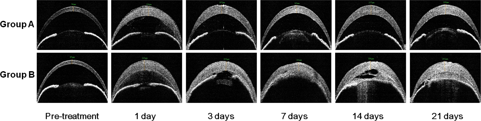
 Figure 2 of
Han, Mol Vis 2013; 19:1222-1230.
Figure 2 of
Han, Mol Vis 2013; 19:1222-1230.