Figure 5. Photoreceptor-like cells in porcine RPE cell culture. A, B are bright field views of porcine RPE cells after 18 h in culture (A) and in a passage 1 culture (B). C-E are a control culture infected with lenti virus control, Lvx-IRES-ZsGreen1, under with bright-field (C), epi-fluorescence for ZsGreen1 (D), or immunostaining for microtubule-associate protein 2 (E). F-I are porcine RPE culture infected with lenti virus Lvx-ngn3-IRES-ZsGreen1 (expressing neurogenin3) under with bright-field
(F), epi-fluorescence for ZsGreen1 (G), immunostaining for Map2 (H), or a merged view (I). Arrows point to ZsGreen1+ cells with a neural morphology. J-M show neuron-like ZsGreen1+ cells (arrow) with aggregate of dark pigment granules (arrowhead) in cultures infected with Lvx-ngn3-IRES-ZsGreen1. N-P are porcine RPE culture infected with Lvx-ngn1-IRES-ZsGreen1 under with bright-field (N), epi-fluorescence for ZsGreen1 (O), immunostaining for recoverin (P). Arrow points to a ZsGreen1+/Recoverin+ cell; arrowhead points to a ZsGreen1¯/Recoverin¯ cell. The scale bars are 25 µm and the one in C applies to all except A, B.
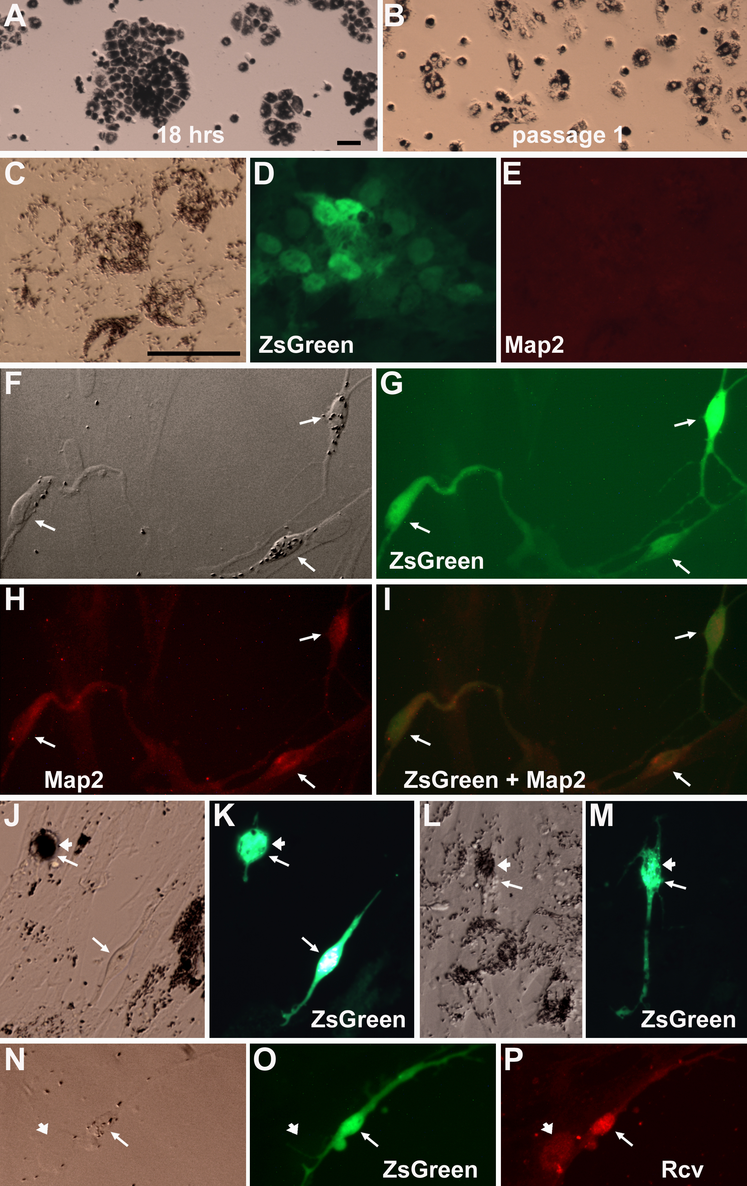
 Figure 5 of
Yan, Mol Vis 2013; 19:1178-1187.
Figure 5 of
Yan, Mol Vis 2013; 19:1178-1187.