Figure 4. Photoreceptor-like cells in a postnatal day 5 mouse RPE cell culture infected with lenti virus Lvx-ngn1-IRES-ZsGreen1. Shown
are bright field (A), epi-fluorescence for ZsGreen1 (B, C, E, G), and immunodetection for microtubule-associate protein 2 (Map2; D), rhodopsin (Rho; F), and red opsin (Red; H). Photoreceptor-like cells are indicated by arrows. Scale bars are 25 μm.
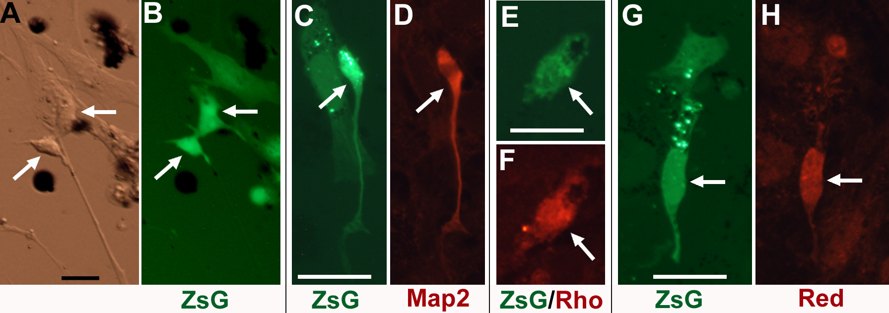
 Figure 4 of
Yan, Mol Vis 2013; 19:1178-1187.
Figure 4 of
Yan, Mol Vis 2013; 19:1178-1187.