Figure 3. Histological assessments of in vivo protective efficacy by contact lenses. A and C: Hematoxylin and eosin (HE) staining of the corneas showed disordered stroma, a thinner epithelium layer, and more apoptotic
vacuoles (indicated by arrows) in the ultraviolet B (UVB) and the Vifilcon A groups after UVB exposure, but not in the groups
with UVB-blocking contact lenses (Etafilcon A and HEMA+MA) or in the blank control group. B: The reduction in the corneal epithelium thickness was significantly prevented in the groups with UVB-blocking contact lenses
(Etafilcon A and HEMA+MA) compared to the UVB group (p<0.001 as determined wt the Wilcoxon-Mann–Whitney test; n=8). D: The corneal stroma showed infiltration of polymorphonuclear (PMN) leukocytes (indicated by arrows) after UVB irradiation,
which was not seen in the eyes with UVB-blocking contact lenses (Etafilcon A and HEMA+MA). E: The infiltrative PMN leukocytes among the five study groups were shown by quantitative analysis (p<0.001 as determined by
the Wilcoxon-Mann–Whitney test; n=5). The scale bar in A-e represents 100 μm, in C-e represents 30 μm, and in D-j represents 15 μm.
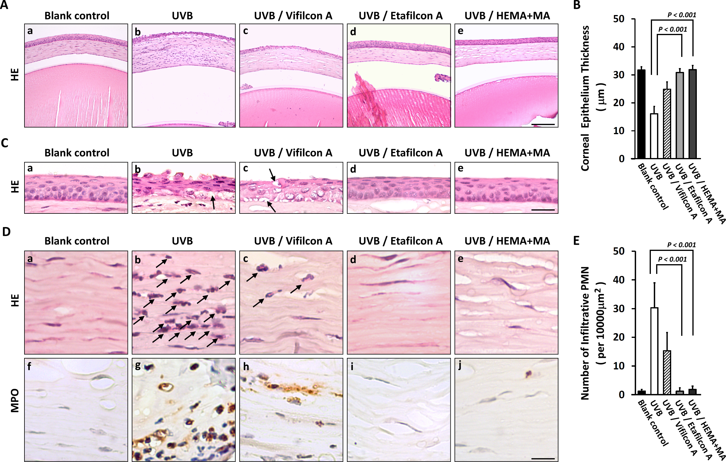
 Figure 3 of
Lin, Mol Vis 2013; 19:1158-1168.
Figure 3 of
Lin, Mol Vis 2013; 19:1158-1168.