Figure 3. In vivo and ex vivo characterization of stromal-derived factor-1 target modification in imidapril-treated animals. Whole blood
(WB) from various animals was collected 12 days following laser-induced choroidal neovascularization (CNV). The lymphocytes
were either stimulated or unstimulated with exogenous SDF-1 and were analyzed using phalloidin staining and FACS analysis.
A significant increase in F-actin polymerization fluorescence intensity was observed following stimulation with SDF-1 (blue
peak) compared with the unstimulated (white peak) lymphocytes from the blood of the vehicle-treated mice, as illustrated by
the two clearly distinct peaks (A: left histogram). SDF-1-stimulated (blue peak) lymphocytes from the blood of imidapril-treated mice exhibited slightly increased
F-actin polymerization fluorescence compared with the unstimulated blood (white peak), as illustrated by the two overlapping
peaks (A: right histogram). B: F-actin polymerization was measured following stimulation with SDF-1. Values are expressed as the mean±standard deviation
(n=5). The asterisk (*) indicates p<0.05. Dip-A represents diprotin-A.
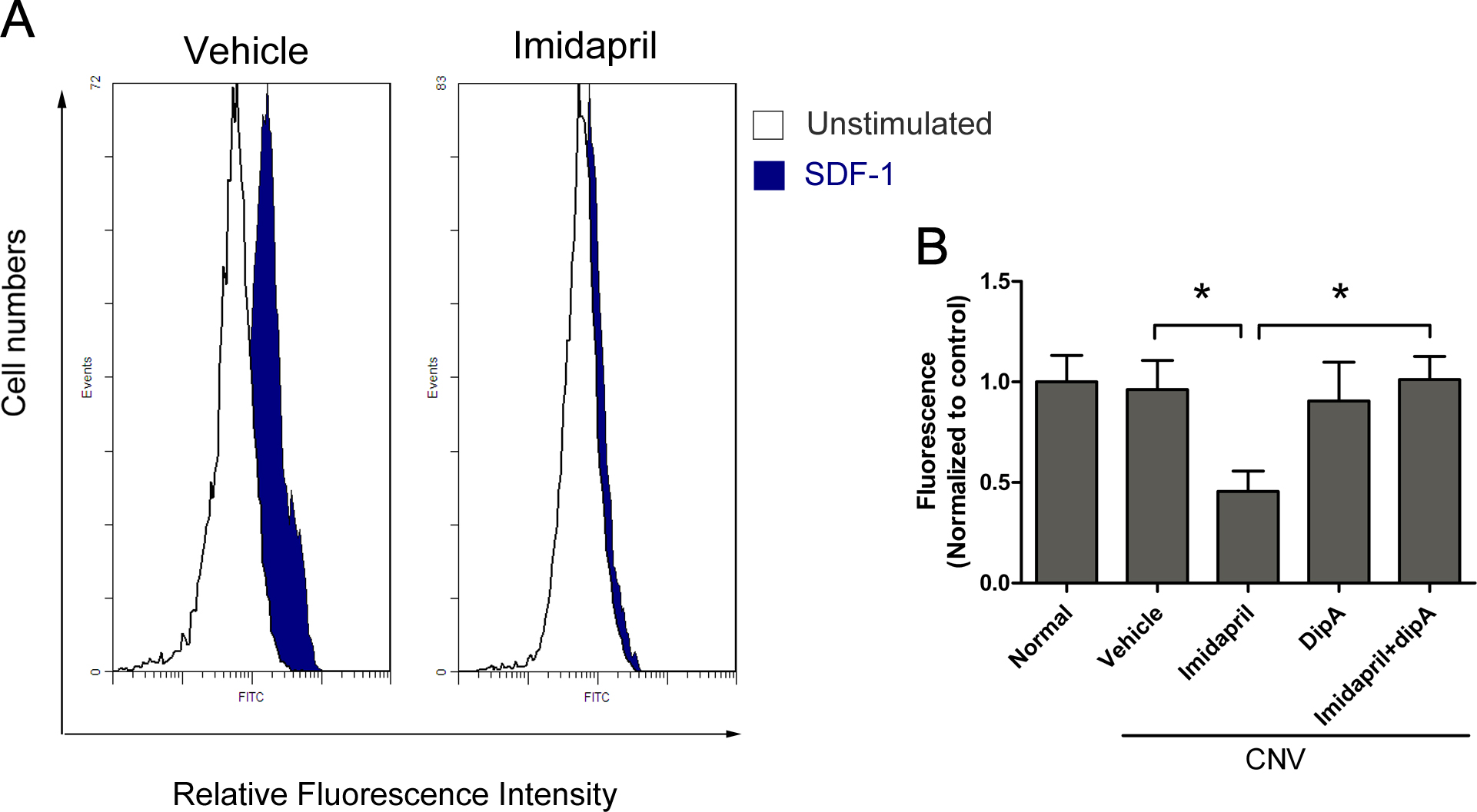
 Figure 3 of
Li, Mol Vis 2013; 19:1107-1121.
Figure 3 of
Li, Mol Vis 2013; 19:1107-1121.