Figure 3. Establishment of mouse corneal endothelial cell (MCEC) cultures. The phase contrast micrographs show the Descemet’s membrane–endothelial
layers stripped from corneas of wild-type mice (A, scale bar = 20 µm; B, scale bar = 50 µm). The morphology of cultured MCECs as observed at early passage 2 (C, scale bar = 20 µm; D, scale bar = 50 µm) and at late passage 7 (E, scale bar = 20 µm; F, scale bar = 50 µm).
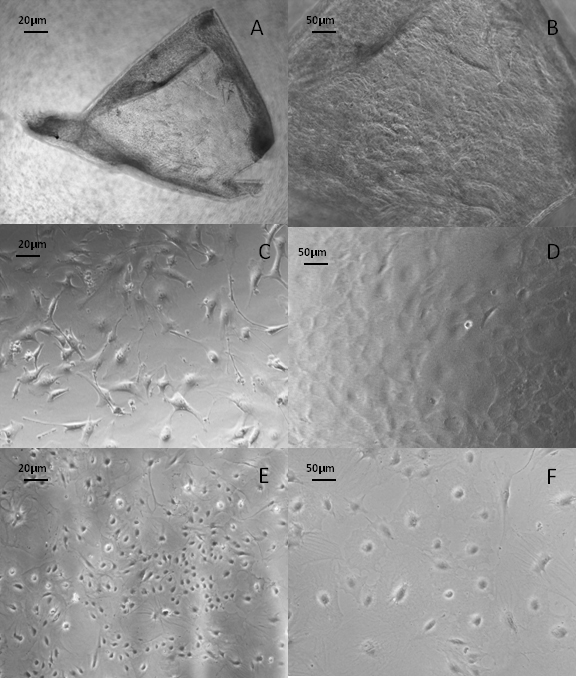
 Figure 3 of
Shei, Mol Vis 2013; 19:1096-1106.
Figure 3 of
Shei, Mol Vis 2013; 19:1096-1106.