Figure 5. Changes in the retina over time can be captured in the same eye. A: An unltraviolet-sensitive (UV) cone that regenerated between 28 days post-lesion (DPL) [A] and 35 DPL [A’] is indicated with white arrows. White chevrons indicate the same cone in both panels, used to match the areas on different
days. B: Faint spots of fluorescence (some examples shown with arrows) suggest that new blue-sensitive cones are beginning to express
sws2 opsin (mCherry) at 10 DPL, and appear smaller and fainter than the surviving cones seen at the lesion edge in upper half
of image. C: The same retina as shown in B, in the GFP channel; the white arrow indicates one or two new UV-sensitive cones beginning to express sws1 opsin. Faint GFP
expression of new UV cones, alongside cones with robust expression, can be viewed in Appendix 3. n=2 fish shown.
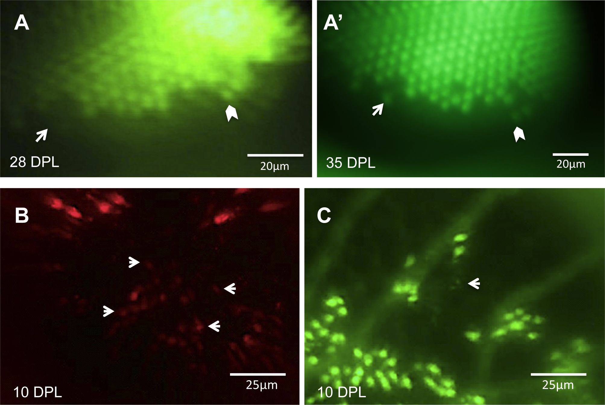
 Figure 5 of
Duval, Mol Vis 2013; 19:1082-1095.
Figure 5 of
Duval, Mol Vis 2013; 19:1082-1095.