Figure 1. Mounting of a custom fundus lens on a fluorescent stereomicroscope allows characterization of individual photoreceptor cells
in vivo. A: An adult anaesthetized zebrafish is shown, positioned on its flank under the objective lens such that its pupil is in the
centre of the field-of-view. The fundus lens is positioned in the light path, centered above and touching the pupil (detailed
in B). A micromanipulator (right side of image) allows precise and constant positioning of the fundus lens but is not required.
B: A view of the custom fundus lens in position for viewing, positioned on the fish eye. The ventral side of the fish is in
view.
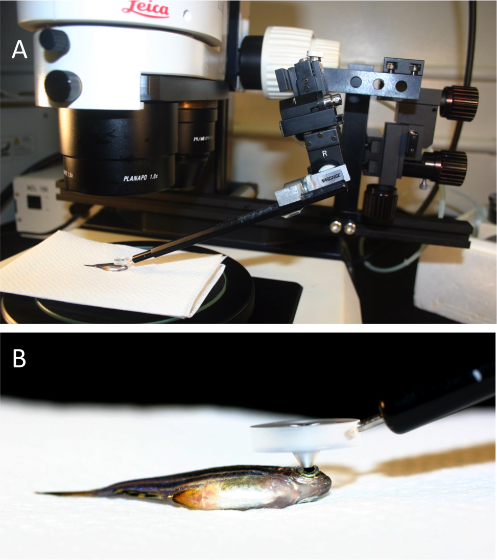
 Figure 1 of
Duval, Mol Vis 2013; 19:1082-1095.
Figure 1 of
Duval, Mol Vis 2013; 19:1082-1095.