Figure 4. O-GlcNAcylated proteins localize to retinal vascular plexus. A: Eye sections from P10 and 7-month-old Ins2Akita/+ mice. B: P12 and P17 oxygen-induced ischemic retinopathy (OIR) wild-type (WT) mice. O-GlcNAcylated proteins labeled with Cy3 (red,
first row), vascular plexus labeled with Cy2 (green, second row) and merge images (third row). Please note the high amount
of O-GlcNAcylated protein colocalization with the retinal vascular plexus in 7-month-old Ins2Akita/+ and P17 OIR eyes (arrowheads). These images are representative of images evaluated in eyes from at least six mice (original
magnification x200).
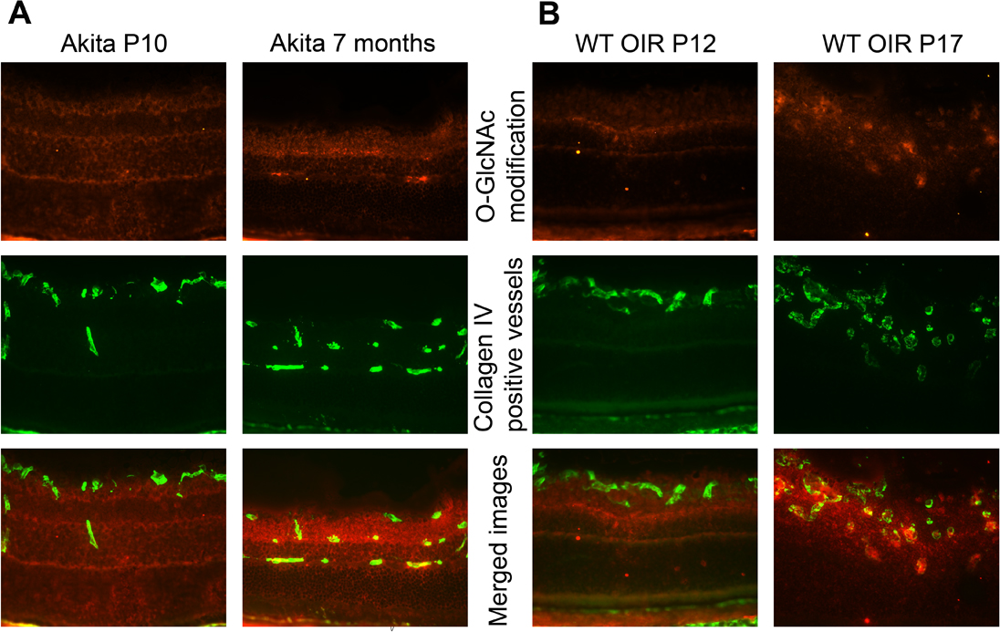
 Figure 4 of
Gurel, Mol Vis 2013; 19:1047-1059.
Figure 4 of
Gurel, Mol Vis 2013; 19:1047-1059.