Figure 1. Increased O-linked-N-acetylglucoseamine modification (O-GlcNAcylation) with increased O-GlcNAc transferase (OGT) and decreased
O-GlcNAcase (OGA) expression during postnatal retinal vascular development and aging. A: Protein lysates (25 µg) from C57BL/6J mouse retinas were analyzed by western blot analysis for O-GlcNAcylated proteins and
the expression of OGT and OGA. B: The β-actin expression was assessed as a loading control and used for normalization and quantification of data, which were
obtained after three different runs (B). RNA expression of OGT and OGA were determined by qPCR and normalized by RpL13A RNA expression in samples. The qPCRs were
performed with three biologic replicates and in triplicate (C). Validation of O-GlcNAc antibody staining of lysates prepared from pericytes (PC) under various glucose conditions. D: The GlcNAc (1 M) competition during primary antibody incubation was used to validate the specificity of the O-GlcNAc RL2
antibody. IgG control was used to validate the existence of the secondary antibody. Mean±SEM; * (p≤0.05), ** (p≤0.01), and
***(p≤0.001) significantly different from P5.
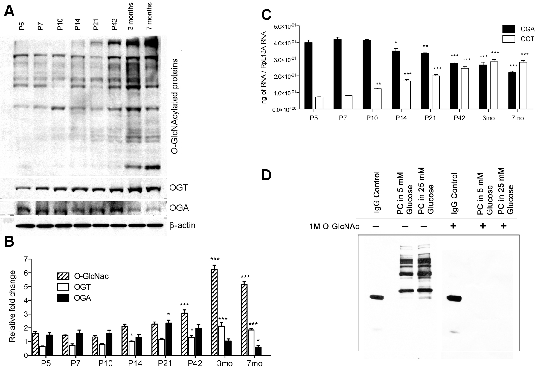
 Figure 1 of
Gurel, Mol Vis 2013; 19:1047-1059.
Figure 1 of
Gurel, Mol Vis 2013; 19:1047-1059.