Figure 3. Macular optical coherence tomography images from a visually normal subject and the proband of this family with cone dystrophy.
A: The macular optical coherence tomography images of the right eye from a normal individual show organization of retinal microstructures
with a well defined photoreceptor inner/outer segment layer and normal thickness (214 μm). B and C: The macular optical coherence tomography images of both eyes from the proband exhibit loss of inner/outer segment layer
and thinning of the retina in the macular area (151 μm of the right eye, 153 μm of the left eye).
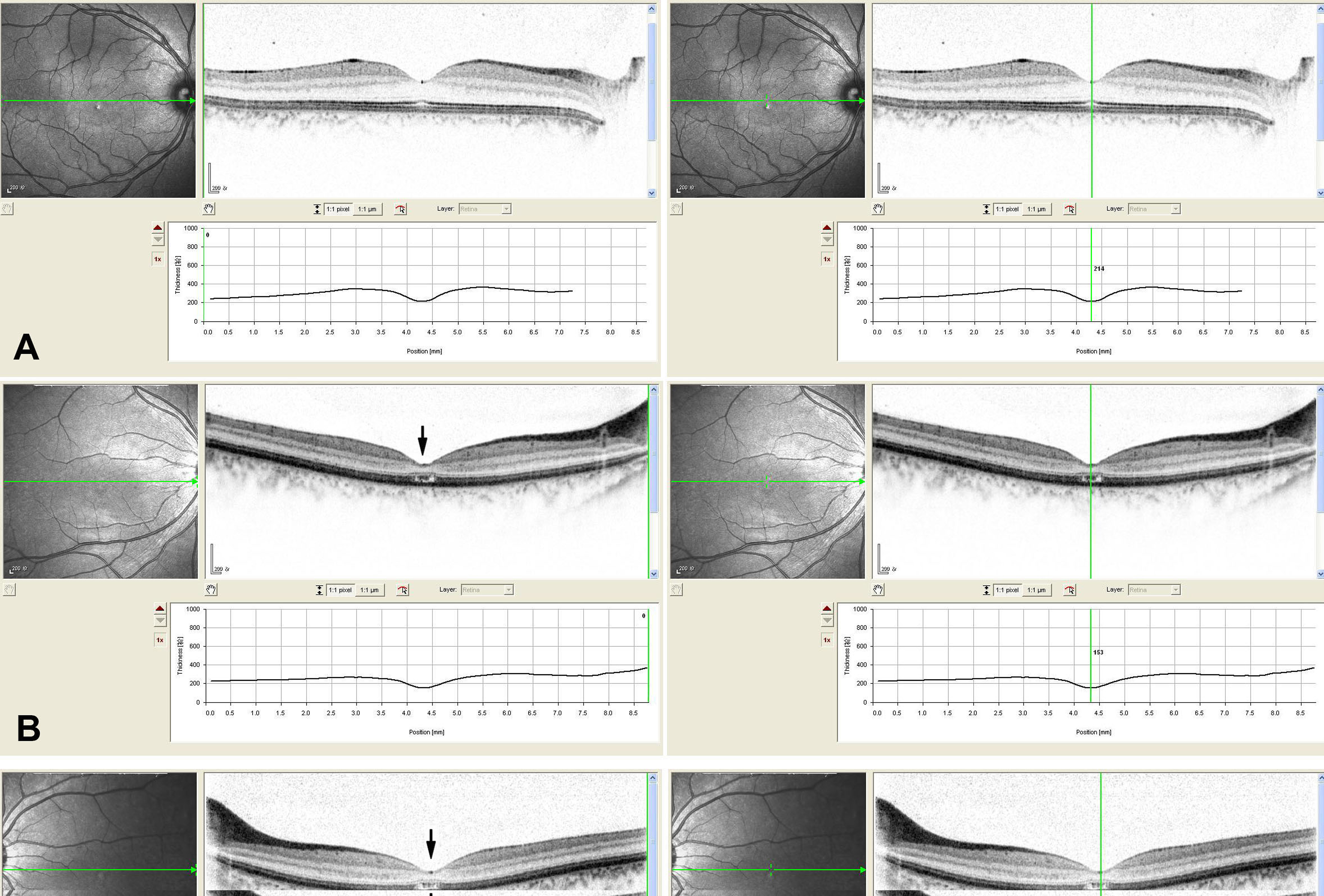
 Figure 3 of
Zhao, Mol Vis 2013; 19:1039-1046.
Figure 3 of
Zhao, Mol Vis 2013; 19:1039-1046.