Figure 4. Characterization and validation of EpCAM antibody conjugation to gold nanoparticles. A: SDS-PAGE shows retardation of EpCAM antibody conjugated AuNP-PEI nanoparticles. EpCAM antibody retardation was observed
along with the increase in EpCAMPEI ratio (0.5 nmol/100 nmol PEI onwards). B: A UV-VIS spectrum shows the differences in the peaks between AuNP-PEI and EpCAM conjugated AuNP-PEI nanoparticles. The UV-VIS
spectrometry analysis of AuNP-PEI-EpAb (549 nm) demonstrated deviation in the absorption spectra from the typical 529nm spectra
of AuNP-PEI.C: Fluorescence microscopy image confirms the uptake of EpCAM conjugated AuNP-PEI nanoparticles. The antibody conjugation was
detected by FITC labeled anti-mouse secondary antibody in the Y79 cells transfected with AuNP-PEI-EpAb nanoparticles (D) compared to cells treated with AuNP-PEI nanoparticles (C).
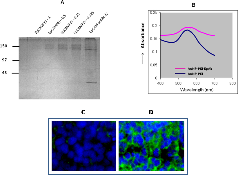
 Figure 4 of
Mitra, Mol Vis 2013; 19:1029-1038.
Figure 4 of
Mitra, Mol Vis 2013; 19:1029-1038.