Figure 3. Effect of actin binding drugs on myocardin-related transcription factor (MRTF-A) migration in rat lens explants. Cells were
immunostained with Alexa Fluor 568 conjugated MRTF-A secondary (red), fluorescein isothiocyanate for alpha smooth muscle actin
(αSMA) (green), and 4', 6-diaminodino-2-phenylindole (DAPI) as a nuclear stain (blue). Treatment with cytochalasin D (CD)
showed cells with bright nuclei as a result of increased nuclear accumulation of MRTF-A (A). In addition, there was an increase in αSMA production by the cells even in the absence of transforming growth factor beta
(TGFβ). B: Latrunculin B (LatB)+ TGFβ cotreated cells showing mostly cytoplasmic and pan-cellular MRTF-A (D) with a decrease in αSMA expression (E). As a whole, the LatB+ TGFβ-treated cells greatly resemble the control cells. The corresponding composite picture with DAPI
(C, F) demonstrates the difference between nuclear and cytoplasmic localization more effectively. Each given scale bar equals 100
μm. Quantification of MRTF-A localization in the rat lens epithelial explants was done using ImageJ (G). Compared to the control, the explant cells showed a significant increase in nuclear MRTF-A and significant decrease in
cytoplasmic MRTF-A with TGFβ (n=7) and CD (n=4) treatment. However, when TGFβ treatment is compared to LatB and TGFβ cotreatment
(n=4), the increase in cytoplasmic MRTF-A and decrease in nuclear MRTF-A are significant (*p<0.05). When the CD and TGFβ treatments
are compared, the change in nuclear MRTF-A is non-significant. Similarly, the difference in nuclear MRTF-A between the control
and LatB-TGFβ cotreatment is insignificant. Image analysis shows αSMA production by the cells (H). Results clearly demonstrate a significant increase *(p<0.05) in αSMA production on TGFβ (n=6) treatment along with a significant
decrease *(p<0.05) in αSMA with LatB- TGFβ (n=4) cotreatment. In addition, there was also an increase in αSMA production with
CD treatment (n=4).
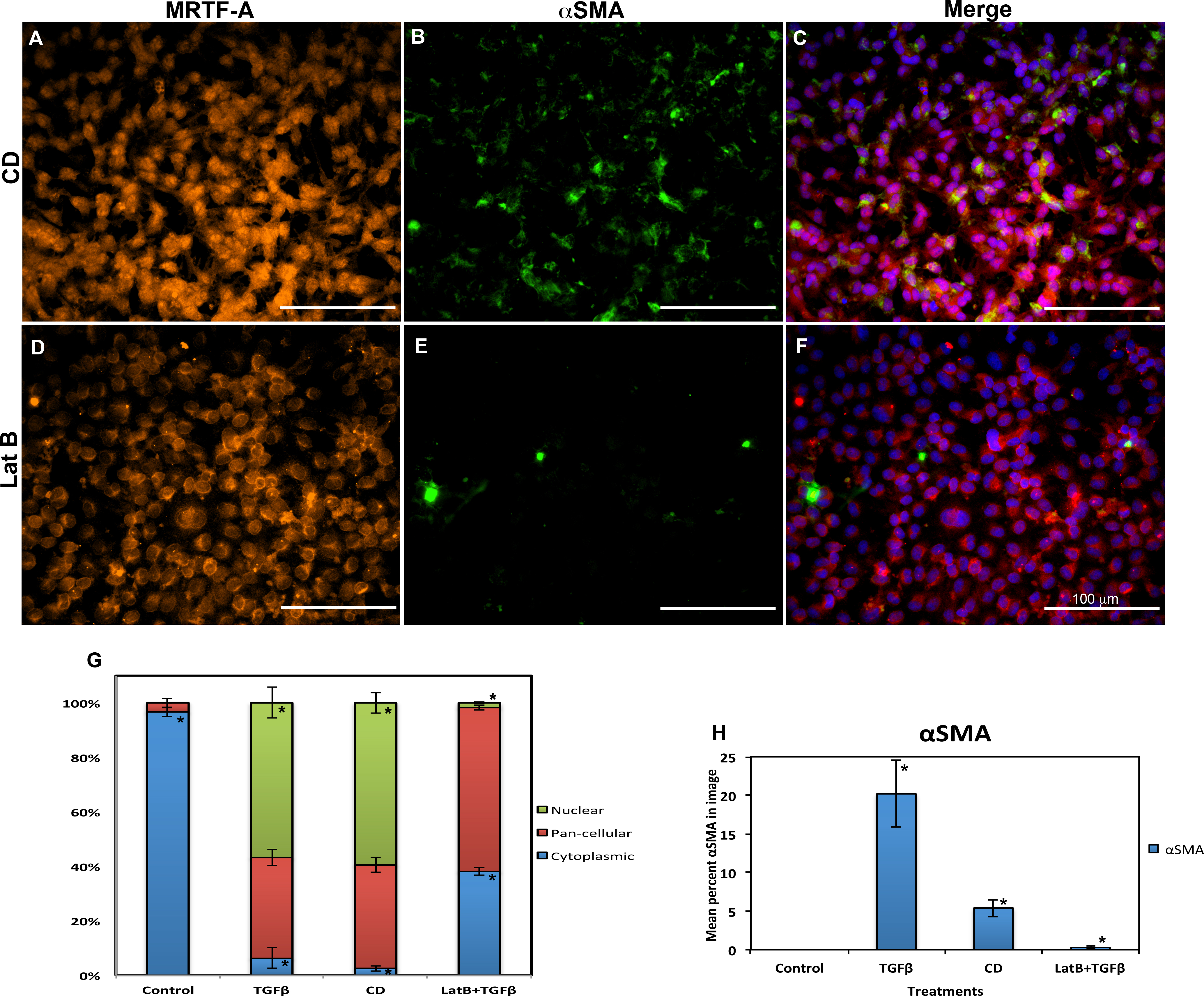
 Figure 3 of
Gupta, Mol Vis 2013; 19:1017-1028.
Figure 3 of
Gupta, Mol Vis 2013; 19:1017-1028.