Figure 1. Localization and quantification of myocardin-related transcription factor-A (MRTF-A) and MRTF-B following transforming growth
factor beta treatment. Cells were treated with 6 ng/ml transforming growth factor beta (TGFβ) for 48 h. The composite pictures
shown here are stained with Alexa Fluor 568 conjugated secondary (red) for MRTF-A and -B and with 4', 6-diaminodino-2-phenylindole
(DAPI) as a nuclear stain (blue). Untreated (control) rat lens explants exhibit cells with primarily cytoplasmic MRTF-A (A) and MRTF-B (B). Following TGFβ treatment, MRTF-A appears to be primarily in the nucleus (C). MRTF-B, however, remains cytoplasmic despite treatment with TGFβ (D). Each scale bar equals 100 μm. Quantification and comparison of intracellular translocation of MRTF-A and MRTF-B in the
rat lens explant cell cultures upon TGFβ treatment were conducted using ImageJ (E). MRTF-A and MRTF-B were primarily cytoplasmic in the control (n=3). A significant increase *(p<0.05) in nuclear MRTF-A and
a significant decrease *(p<0.05) in cytoplasmic MRTF-A were observed after TGFβ treatment (n=7). However, in the case of explants
stained for MRTF-B, no significant change was observed in the intracellular localization following TGFβ treatment (n=3).
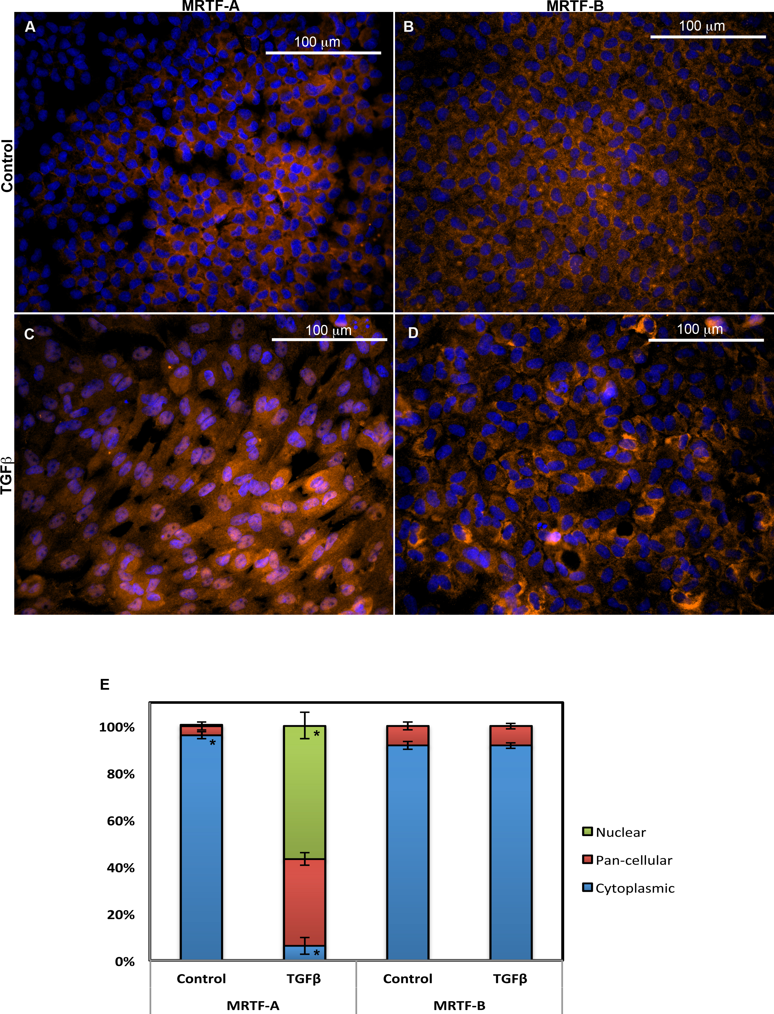
 Figure 1 of
Gupta, Mol Vis 2013; 19:1017-1028.
Figure 1 of
Gupta, Mol Vis 2013; 19:1017-1028.