Figure 2. HIF-1α inhibition does not affect VEGF expression. A: western blot analysis of HIF-1α expression in HLE-B3 cells treated with topotecan. Cell lysates were collected from cells
treated with 500 nM topotecan in 0.01% DMSO after 8 h of hypoxic incubation. Control cells were mock treated with 0.01% DMSO
and maintained in hypoxia as the topotecan-treated cells. Twenty μg protein/lane of cell lysates were analyzed with western
blot analysis, and lane loading was normalized using a 1:1,000 dilution of rabbit anti-pan-actin antibody. Topotecan inhibited
the expression of HIF-1α (1:1,000 dilution of rabbit anti- HIF-1α antibody) while a compensatory increase in HIF-2α was noted
(1:1,000 dilution of rabbit anti- HIF-2α antibody). B, C: Densitometry analysis of HIF-1α and HIF-2α, respectively. D: Effect of HIF-1α inhibition on VEGF synthesis in hypoxia. HLE-B3 cells were cultured in 25 cm2 flasks with 20% FBS and switched to serum-free media 24 h before the experiment. The cells were incubated with 3 ml of serum-free
media containing 500 nM topotecan or 0.01% DMSO for 8 h of hypoxic exposure. Cell-free supernatants were collected in triplicate
at the end of hypoxic incubation and analyzed for VEGF levels with ELISA. There was no significant difference in the VEGF
levels between the topotecan-treated cells and control cells. (A Student t test was performed to compare the VEGF levels between control and treated sample, and the p value was greater than 0.05.)
E: HIF-2α protein from HLE-B3 control cells compared with a standard lysate prepared from human embryonic kidney cells (HEK)
293 (Novus Biologicals, Litton, CO). The HIF-2α found in the standard lysate migrated at about 97 kDa. The HIF-2 α from HLE-B3
cells was about 80 kDa.
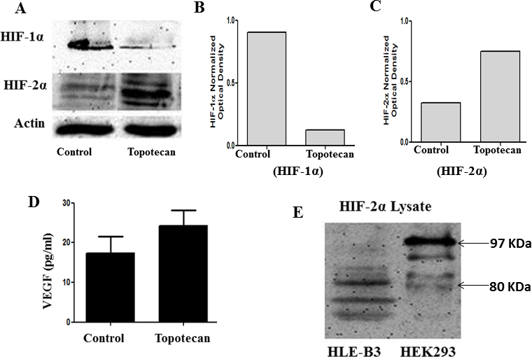
 Figure 2 of
Neelam, Mol Vis 2013; 19:1-15.
Figure 2 of
Neelam, Mol Vis 2013; 19:1-15.