Figure 1. Sustained and cumulative expression of VEGF in hypoxia and atmospheric oxygen. Detection of VEGF levels in hypoxia with ELISA.
HLE-B3 cells were cultured in 25 cm2 flasks with 20% FBS and switched to serum-free media 24 h before the experiment. The cells were incubated with 3 ml of serum-free
media in hypoxia (1% oxygen) or remained in atmospheric oxygen (about 21% oxygen) for up to 72 h. Cell-free supernatants were
collected in triplicate at 8, 24, 48, and 72 h and analyzed with ELISA to detect the VEGF levels. VEGF consistently accumulated
throughout the 72 h incubation period regardless of whether the cells were maintained in hypoxia or atmospheric oxygen (p<0.05).
A Student t test was performed to compare the VEGF levels between hypoxia and normoxia. Significantly higher levels of VEGF were detected
at all-time points beyond the initial 8h point in hypoxia compared with atmospheric oxygen. Error bars are not shown at time
points because the symbol is larger than the error bar.
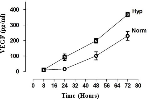
 Figure 1 of
Neelam, Mol Vis 2013; 19:1-15.
Figure 1 of
Neelam, Mol Vis 2013; 19:1-15.