Figure 6. Absence of rhodopsin and
scarce presence of recoverin-positive human cells in grafts at 3
weeks after transplantation. There were no rhodopsin-positive
cells in neural grafts at this time point, although rhodopsin
staining was present in the outer segments of the host retina as
expected. Only few recoverin-positive cells were identified in
subretinal grafts at this time point. The abbreviations used in
this figure were the following: RGC, retinal ganglion cells;
INL, inner nuclear layer; ONL, outer nuclear layer. The scale
bar used in this figure represents 50 μm.
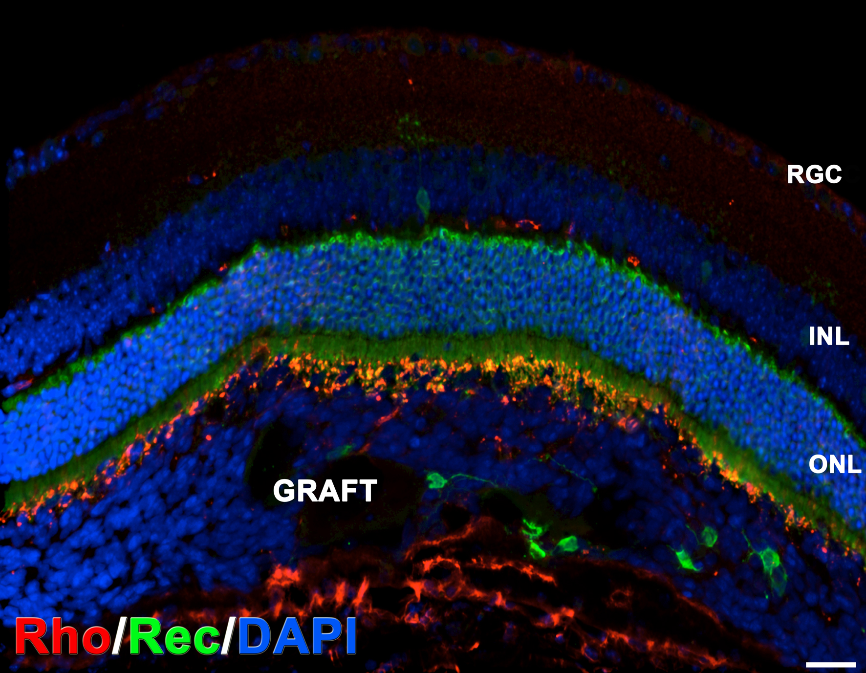
 Figure 6
of Hambright, Mol Vis 2012; 18:920-936.
Figure 6
of Hambright, Mol Vis 2012; 18:920-936.