Figure 5. Microglia accumulation in a
subretinal graft with damaged retinal pigment
epithelium/choroid. This is a typical staining pattern (A,
B) observed in grafts where a needle penetrated retinal
pigment epithelium (RPE) and disrupted choroid (Chr) vasculature
(solid white arrows in B), leading to the rupture of the
retinal–blood barrier and exposure of xenogenic (human) graft to
the host’s immune system. By 3 weeks after subretinal
transplantation, there are typically no surviving human neurons
in such grafts, yet some human nuclei-positive immunoreactivity
occasionally may be found. Solid white arrowheads point to the
accumulation of ionized calcium binding adaptor molecule 1
(Iba-1) staining where the grafted cells were placed. The area
of the main image displayed in the inset in panel B is
indicated with an asterisk (*). The inset shows several
Iba-1-positive cells with a morphology typical for activated
microglia. The scale bar used in panel B is 50 μm.
Double asterisk (**) in panel B indicates the area,
enlarged in panels C and D. This is the host
photoreceptor layer with high microglial activity, where human
retinal progenitors were earlier grafted but did not survive.
Microglial processes are shown with double white arrowheads. The
following abbreviations were used in these panels: ONL – outer
nuclear layer, INL, inner nuclear layer, RGC- retinal ganglion
cells, DAPI – 4', 6-diamidino-2-phenylindole.
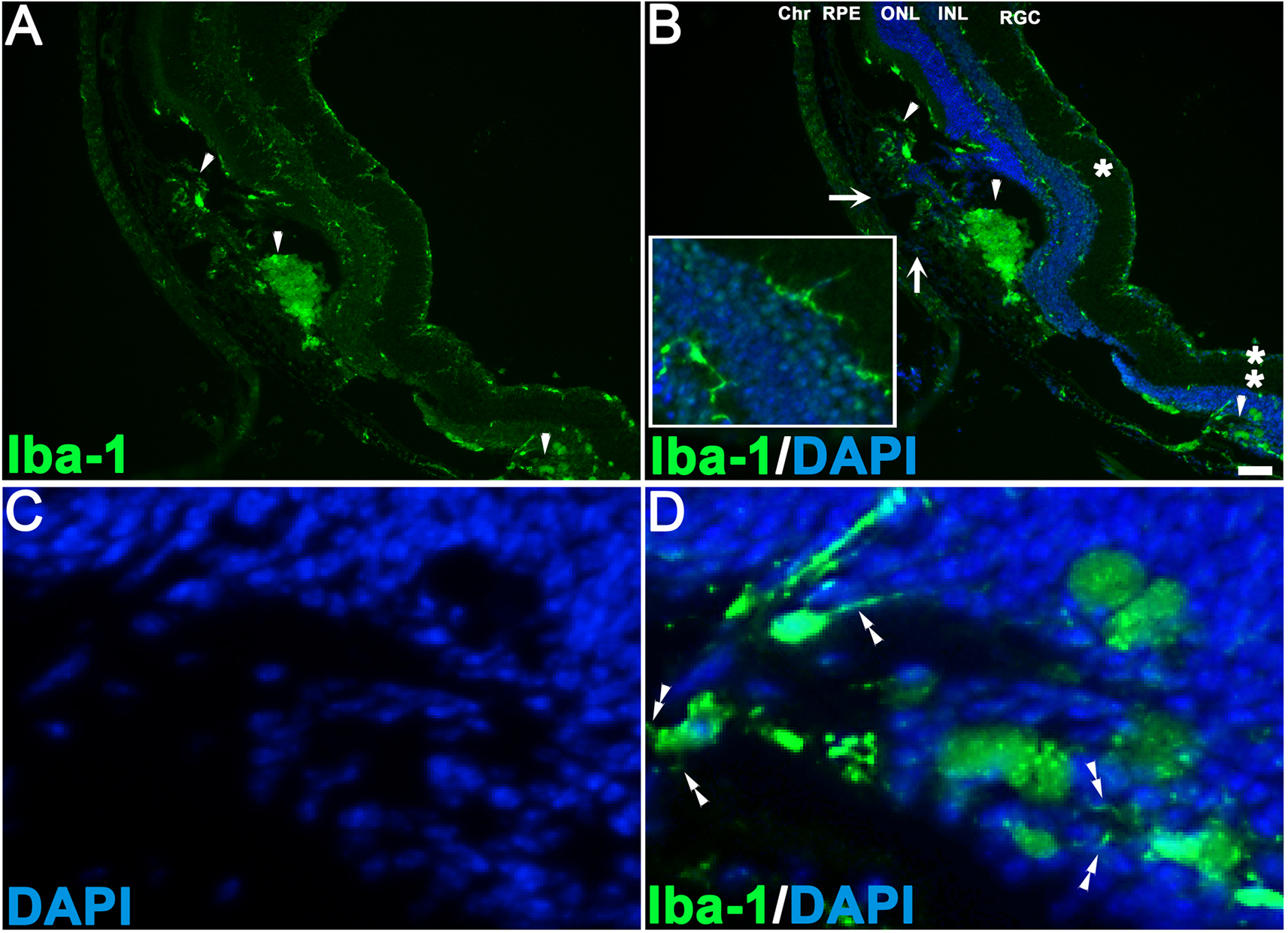
 Figure 5
of Hambright, Mol Vis 2012; 18:920-936.
Figure 5
of Hambright, Mol Vis 2012; 18:920-936.