Figure 4. Glial fibrillary acidic
protein (GFAP) activation in the host retina around the grafting
site 3 weeks after transplantation. GFAP activation (A)
was found in all examined cases where the subretinal grafts were
found, regardless of whether the grafts survived or not. In the
case shown, the release of human nuclei –positive (HNu [+])
immunoreactivity was found in the grafting site outside of the
nuclei, indicating the initial stage of graft destruction (B,
C). Inset in C shows a low-power image of the
same graft from which the main panel was derived. Panel D
displays the staining of nuclei of both human and mouse cells
with 4', 6-diamidino-2-phenylindole (DAPI). The scale bar used
in panels A-D is 50 μm. The outer nuclear layer
(ONL) around the grafting site was damaged by the needle. The
asterisk indicates the area shown in the inset. Abbreviations
used in this legend are the following: INL, inner nuclear layer;
RGC, retinal ganglion cell (layer).
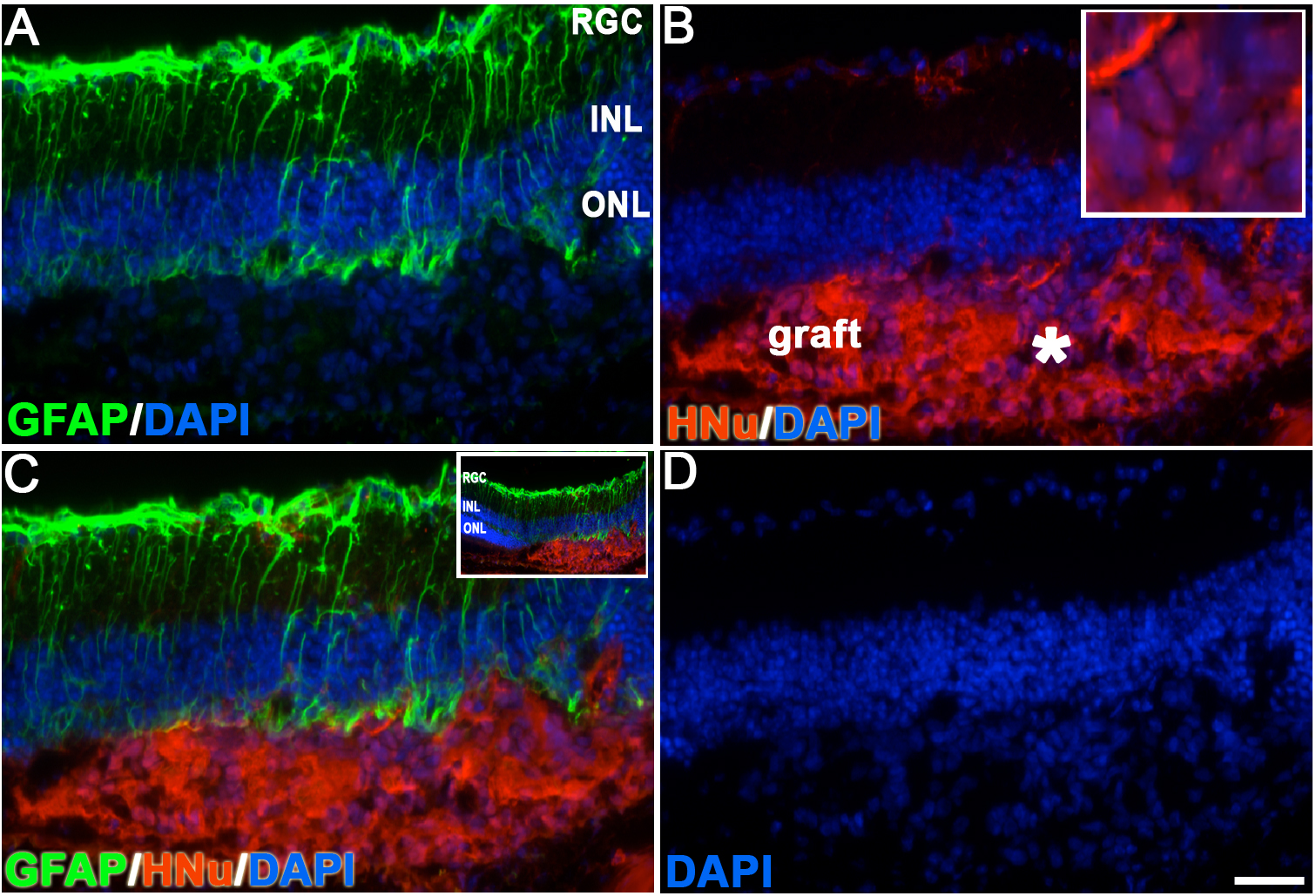
 Figure 4
of Hambright, Mol Vis 2012; 18:920-936.
Figure 4
of Hambright, Mol Vis 2012; 18:920-936.