Figure 2. Limited retinal integration but substantial maturation of human embryonic stem cell-derived retinal progenitor cells in subretinal
grafts. Immunostaining for A; doublecortin (DCX) and human nuclei (HNu), B; neuronal class III β-tubulin (Tuj1 epitope) and HNu, and C; Photoreceptor (PR) marker recoverin and HNu in subretinal grafts. The asterisks indicate the areas shown in insets. Note
the border (indicated with closed white arrowheads) demarcating the outer nuclear layer (ONL) and the grafts in A and B showing the location of the outer limiting membrane (OLM). Subretinal grafts consisted of mostly early neuronal cells (~76%),
as shown in D. D represents grouped data for both human embryonic stem cell (hESC) lines. Error bars indicate the standard error of the mean
(SEM), (the means did not differ significantly between the two hESC lines by an unpaired Student’s t-test). The Tuj1 marker is still heavily present in subretinal grafts at 3 months (E-H); however, the cells fail to migrate into the host’s ONL and remain in the subretinal space (E, H) unless the neural retina (NR) is damaged by injection (F). As in A and B, the host’s OLM in the 3-month grafts presents a barrier for subretinally grafted cells to migrate into the host’s ONL (G, shown with closed white arrowheads), although many grafts spread over a large subretinal area (also in G). H: Cells from the epiretinal grafts integrate into the host’s retinal ganglion cell (RGC) layer, inner plexiform layer, and
inner nuclear layer (INL). I-L: Mature immunophenotypes, such as recoverin (I, J), synaptophysin (K), and rhodopsin (L), are observed in these grafts. In I and J, subretinal grafts predominantly display recoverin-positive (an early photoreceptor) immunophenotype (shown in z-sections, see insets in I and J). The asterisks point to areas within the main images shown in these insets. In I and J we compare the integration of early hESC-derived photoreceptors into the host ONL in intact, nondamaged retinas (I) versus that, where retinas were damaged by the injection needle (J). Compare I, no integration of HNu [+] cells in the host’s ONL can be seen, versus J, where Rec (recoverin) [+], HNu [+] PRs are found embedded into the host’s ONL (indicated with two asterisks). Also see the
respective inset (** in J). In K note sparse human synaptic boutons (shown with white arrows and enlarged in inset) stained with human-specific synaptic marker
synaptophysin (Syn) found both on graft- (shown in inset) and host-specific neurons. The asterisks point to areas within the
main images shown in the insets. The main images in the panel are confocal images. The insets show confocal z-stack analysis of selected areas done with virtual resectioning along the x and y planes. Scale bars: 20 μm (A, B, E, F, I, J, L); 50 μm (C, G, H); 10 μm (K).
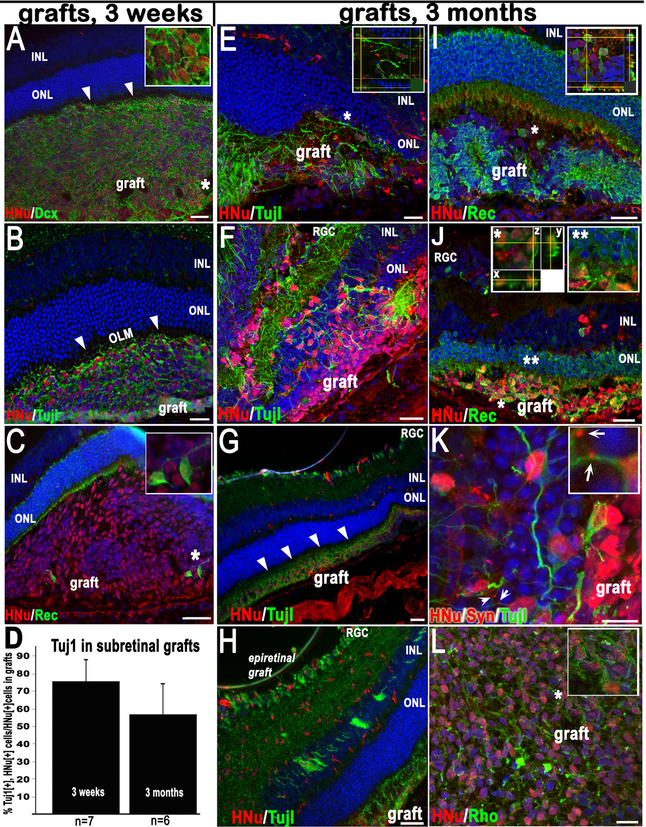
 Figure 2 of
Hambright, Mol Vis 2012; 18:920-936.
Figure 2 of
Hambright, Mol Vis 2012; 18:920-936.