Figure 3. Cell viability and heme
oxygenase-1 (HO-1) expression in human retinal pigment
epithelial cells between curcumin pretreatment and control
dimethyl sulfoxide (DMSO) groups with various H2O2
concentrations. A: Cell viability measured with
3-(4,5-dimethylthiazol-2-yl)-2,5-diphenyltetrazolium bromide
(MTT) assay. Human retinal pigment epithelial cells (ARPE-19)
cells pretreated with 15 μM curcumin showed more cell viability
than those from the control DMSO group. The difference was
highest in 1000 μM H2O2. *p<0.05 . The
percentage of live cells among the total cells was calculated
from three separate experiments. B: HO-1 protein
expression was measured with western blot analysis. The plus
sign means pretreatment with 15 μM curcumin. HO-1 expression was
higher in the curcumin-pretreated group under treatment of H2O2
compared to the control group. β-actin served as the standard.
Results were quantified with densitometry. The image for HO-1
was shown from more than three separate experiments.
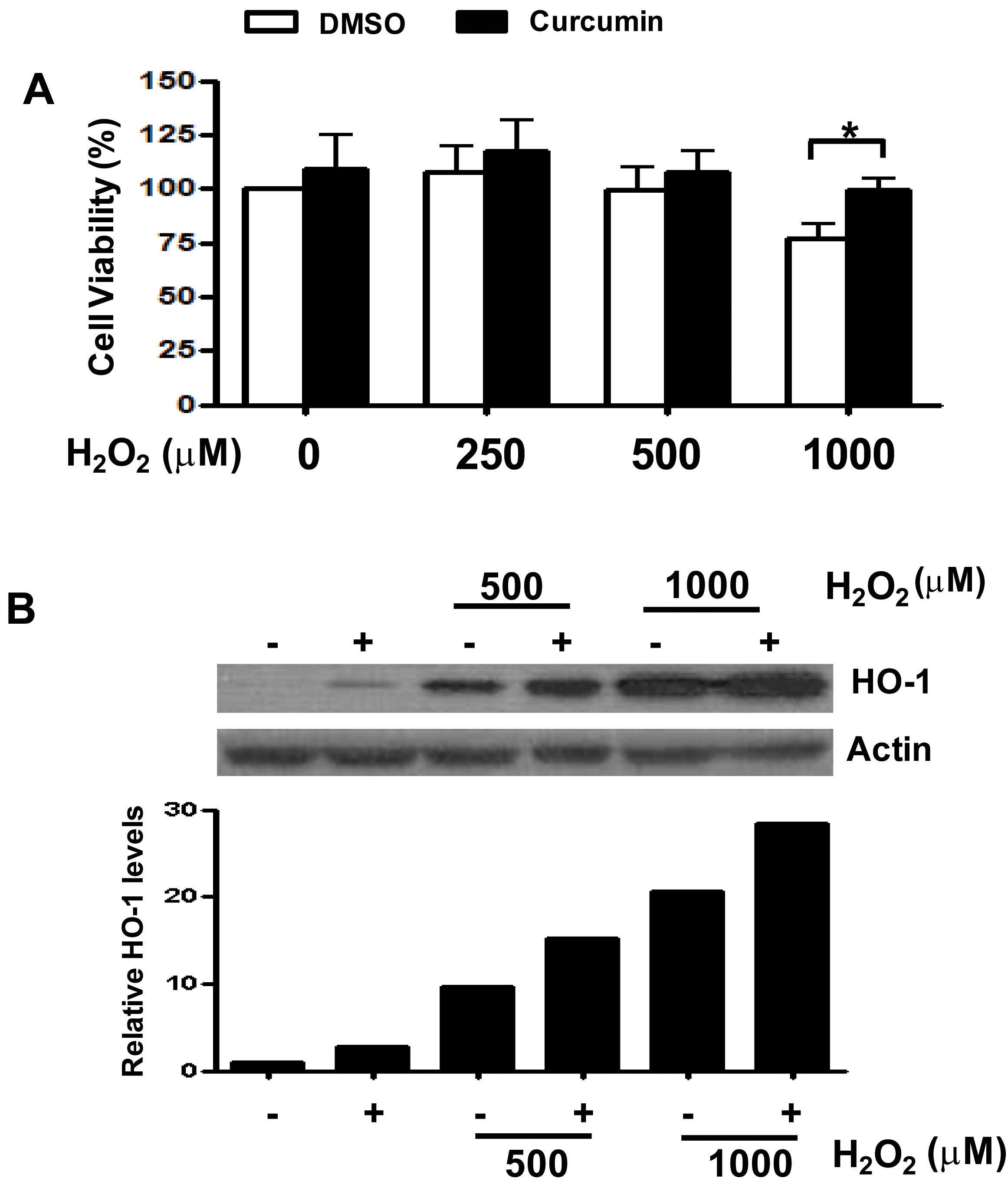
 Figure 3
of Woo, Mol Vis 2012; 18:901-908.
Figure 3
of Woo, Mol Vis 2012; 18:901-908.