Figure 5. Immunofluorescence analysis
showed alpha smooth muscle actin (α-SMA; A), glial
acidic fibrillary protein (GFAP; B), glutamine synthase
(GS; E), and retinal pigment epithelia Protein 65
(RPE-65; F) in epiretinal membranes (EMs) of
proliferative vitreoretinopathy (PVR) model eyes, indicating
fibroblast cells, Müller cells, astroglial cells, and RPE cells
involved in the process of PVR. Hoechst 33342 for nucleic acid
stained alone (C, G). D is the merged
picture of A-C, H the merged picture of E-G
(a triple staining). Arrow shows EM.
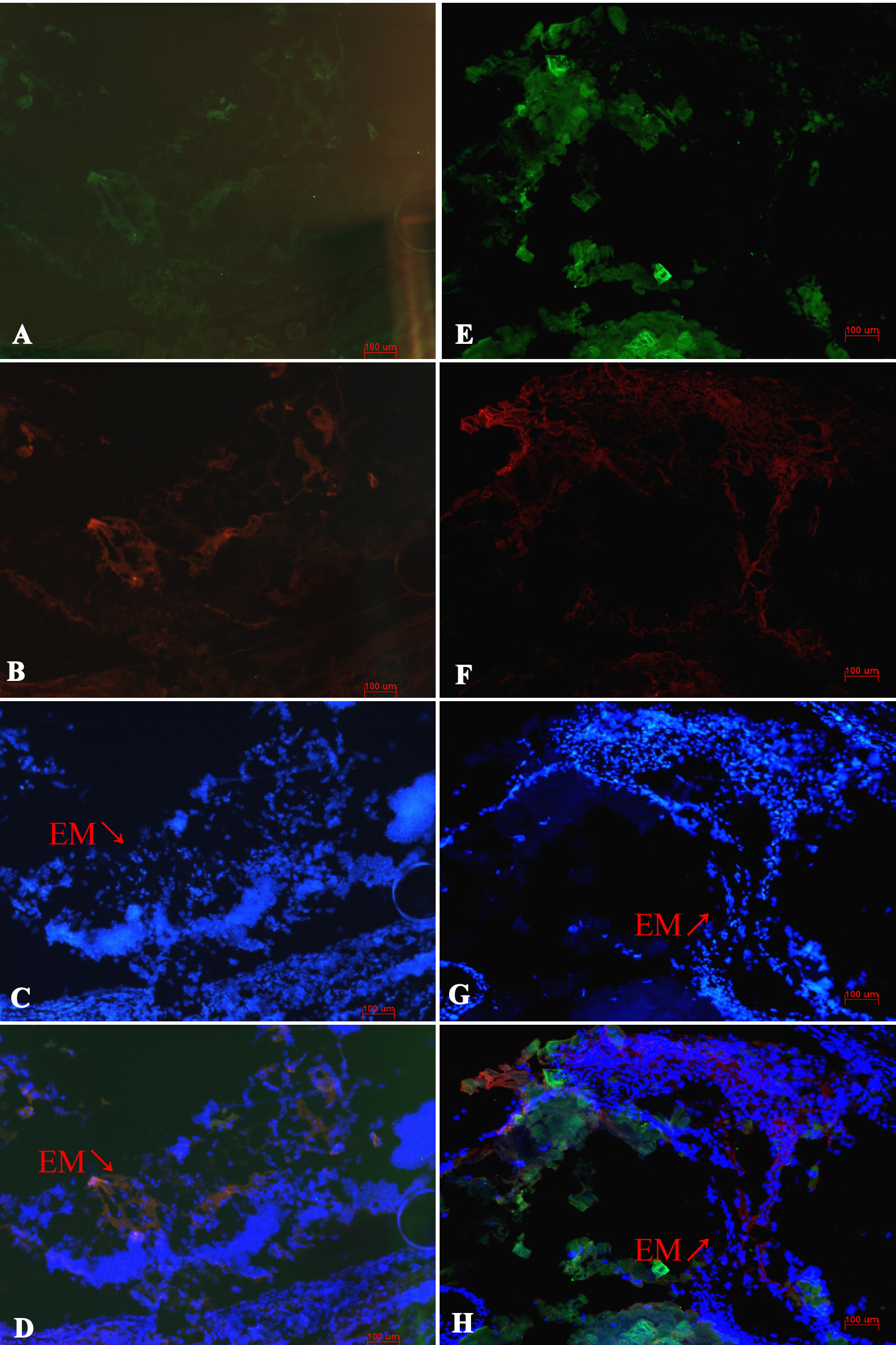
 Figure 5
of Tan, Mol Vis 2012; 18:887-900.
Figure 5
of Tan, Mol Vis 2012; 18:887-900.