Figure 4. Dispase-injected mice
developed cardinal features of proliferative vitreoretinopathy
(PVR). A: Ocular fundi of dispase-injected eye and
saline-injected control eye. Arrow shows PVR membranes. B:
Hematoxylin and eosin (H&E) staining of dispase-injected PVR
eye and saline-injected control eye. Arrow shows proliferative
epiretinal membranes (EM). C: PVR percentages at 1, 2,
4, 6, and 8-week time points after dispase injection.
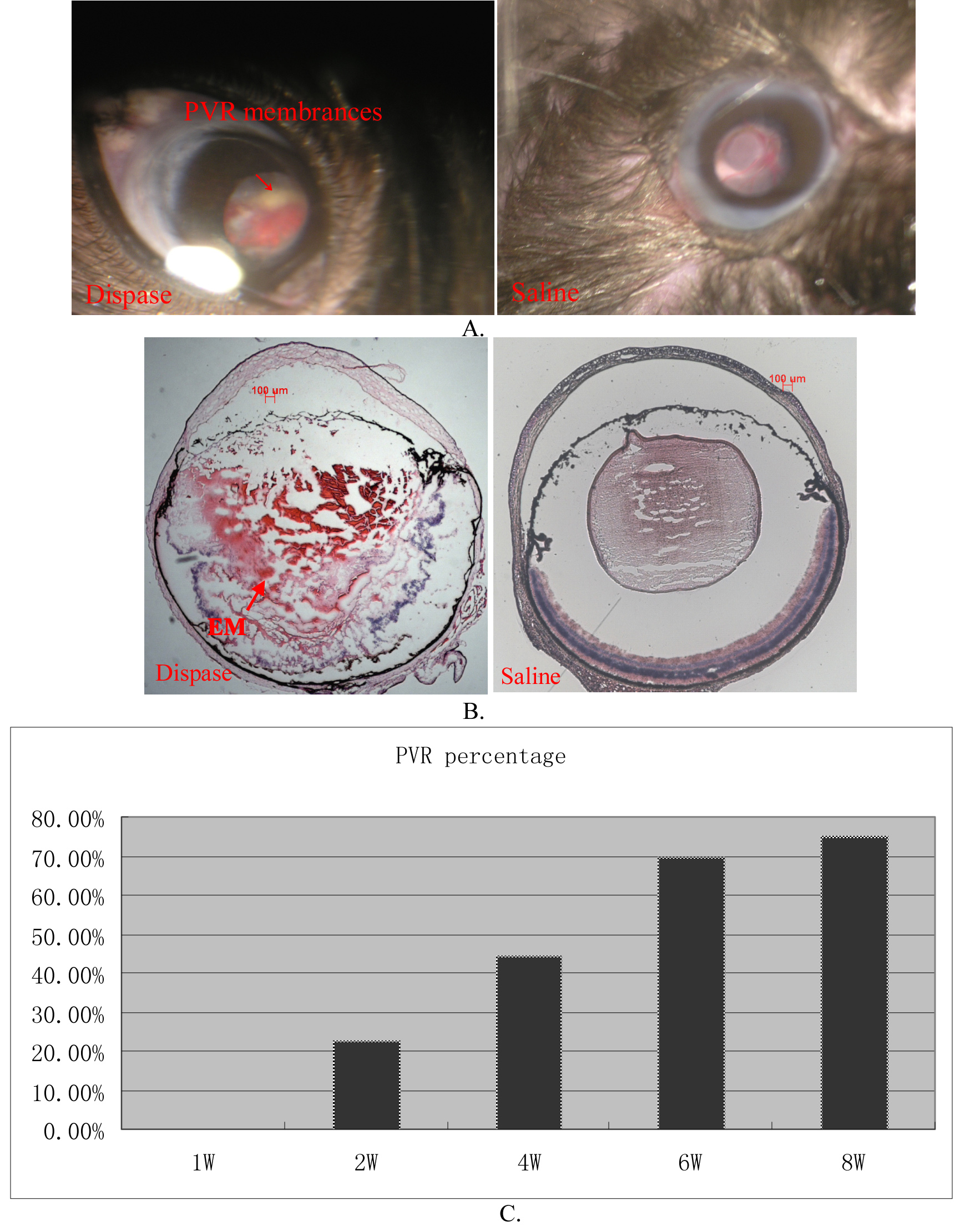
 Figure 4
of Tan, Mol Vis 2012; 18:887-900.
Figure 4
of Tan, Mol Vis 2012; 18:887-900.