Figure 3. Immunofluorescence analysis
showed no cluster of differentiation (CD)3+ labeled T cells,
F4/80+ labeled macrophages, or CD56+ labeled natural killer (NK)
cells involved in dispase-injected eyes at 48 h time point (A,
B). Bluish cells stained with Hoechst 33342 (scale bar
100 μm).
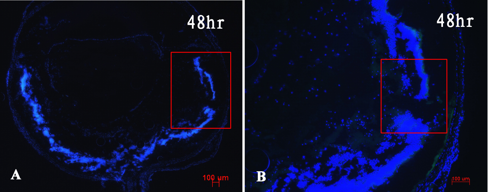
 Figure 3
of Tan, Mol Vis 2012; 18:887-900.
Figure 3
of Tan, Mol Vis 2012; 18:887-900.