Figure 2 of
Tan, Mol Vis 2012; 18:887-900.
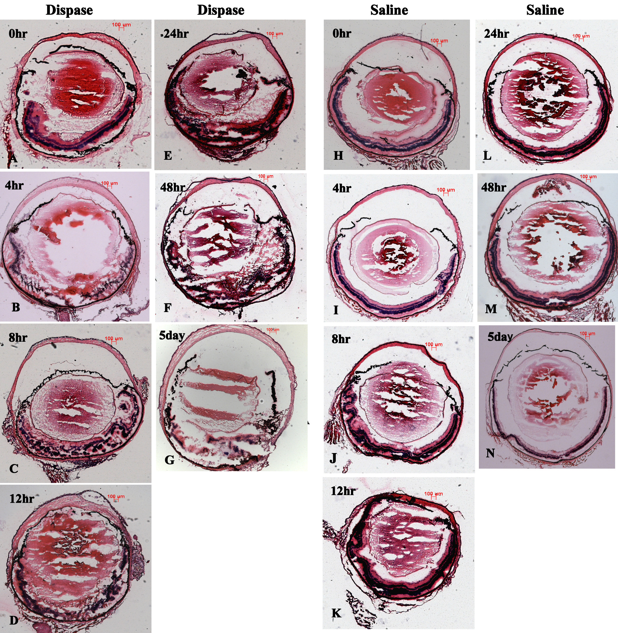
Figure 2.
Ocular morphology during early inflammatory infiltration phase showed retinal structure damaged, in diapase-injected eyes but relatively intact in saline-injected eyes (H&E staining, scale bar 100 μm;
A
-
K
).
 Figure 2
of Tan, Mol Vis 2012; 18:887-900.
Figure 2
of Tan, Mol Vis 2012; 18:887-900.