Figure 6. Intravitreal injection of
labeled siRNA penetrates the retina. A: Confocal
microscopy revealed that the scrambled siRNA covalently bound to
Cy3 (Red) was distributed throughout the layers of the retina
when injected into the eyes of control rats. The label was
distributed throughout the retina from the ganglion cell layer
(GCL) to the photoreceptor layer (PRL). Nuclei were stained with
To-Pro Blue. B: A photomicrograph illustrates panel A
with To-Pro Blue removed. Note the distribution of Cy3 labeled
siRNA throughout the layers of the retina. C: In
contrast, the retinas of rats injected with unlabeled scrambled
siRNA did not contain labeled particles, indicating that the
label was not due to autofluorescence or artifacts from the
injection. Bar=30 um. D: Illustrates higher
magnification of inset depicted with dotted rectangle in A.
Bar=15 um. IPL=inner plexiform layer, INL=inner nuclear layer.
n=3/group.
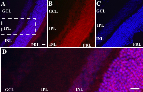
 Figure 6
of Winkler, Mol Vis 2012; 18:874-886.
Figure 6
of Winkler, Mol Vis 2012; 18:874-886.