Figure 3. Connective tissue growth
factor (CTGF) was detected in vimentin-positive Müller cells in
the retina of diabetic rats. A: Müller cells labeled
with filament protein vimentin throughout the diabetic retina. B:
CTGF staining in the 12-week diabetic retina is also seen
throughout the retina. C: Merge photomicrograph for
vimentin and CTGF shows that CTGF colocalizes with vimentin in
the diabetic retina. Bar=60 um. D: High magnification of
the area indicated with a dotted rectangle in C. Bar=5
um. V=Vitreous, ONL=Outer Nuclear Layer, S=Sclera.
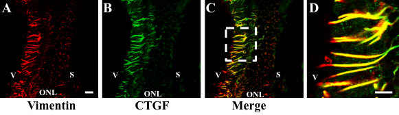
 Figure 3
of Winkler, Mol Vis 2012; 18:874-886.
Figure 3
of Winkler, Mol Vis 2012; 18:874-886.