Figure 7. Measurement of nuclear factor-kappa B (NF-κB) pathway proteins by western blotting in corneas. A: Total lysates from murine corneal tissue lysates were analyzed by western blotting for their level of NF-κB activation by
detecting phosphorylated NF-κB (P-NF-κB), and total NF-κB protein. As shown by the analysis results (B), HIF-1α RNAi-A significantly down-regulated NF-κB phosphorylation expression. Three independent experiments were conducted,
and data were shown as mean±SD **p<0.01 as compared with the Vehicle-RNAi group. C: Immunohistochemistry staining of P-NF-κB in corneas with CL. The arrows indicated the positive staining of P-NF-κB in the
corneal epithelium of each group. P-NF-κB was located to the nucleus. Magnification: 400×.
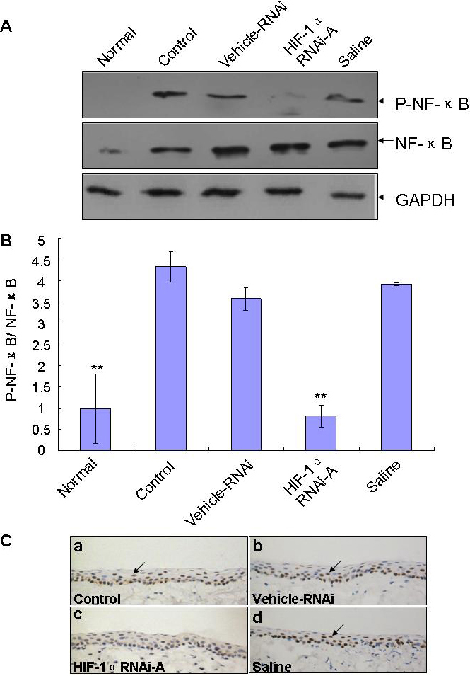
 Figure 7 of
Chen, Mol Vis 2012; 18:864-873.
Figure 7 of
Chen, Mol Vis 2012; 18:864-873.