Figure 6. Western blot analysis of cytochrome c and cleaved caspase-3 protein. At 48 h after transfection, the cells were harvested
and processed for western blotting. β-Actin was used as the internal control. The immunoblot was a representative of three
independent experiments. The expression of cytochrome c and the activity of cleaved caspase-3 were greatly improved (p<0.05)
in the PAMAM group and liposome group, compared with the negative control group and the empty control group.
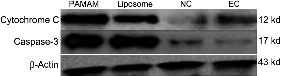
 Figure 6 of
Wu, Mol Vis 2012; 18:74-80.
Figure 6 of
Wu, Mol Vis 2012; 18:74-80.