Figure 2. The distribution of
high-mobility group box protein 1 (HMGB1) in retinal ganglion
cell (RGC)-5 cells changed following treatment with advanced
glycation end products–BSA (AGE-BSA). A:
Immunofluorescence photographs (400× magnification) show that
HMGB1 appears in the cytoplasm (arrow) of RGC-5 cells at 3 h
after treatment with 200 μg/ml of AGE-BSA, while HMGB1 remains
in the nucleus of RGC-5 cells treated with either BSA (200
μg/ml) or PBS. The left column shows the distribution of HMGB1
protein with green fluorescence. The central column shows the
nucleus stained with 4',6-diamidino-2-phenylindole (DAPI; blue),
and the right column shows merged pictures. B:
Subcellular fractionation of proteins shows a significant
increase of HMGB1 levels in the cytosol of RGC-5 cells after
incubation with AGE-BSA for 3 h (n=4, p<0.05, BSA-treated
cells were used as control). The level of HMGB1 in the nucleus
was not different between groups over time.
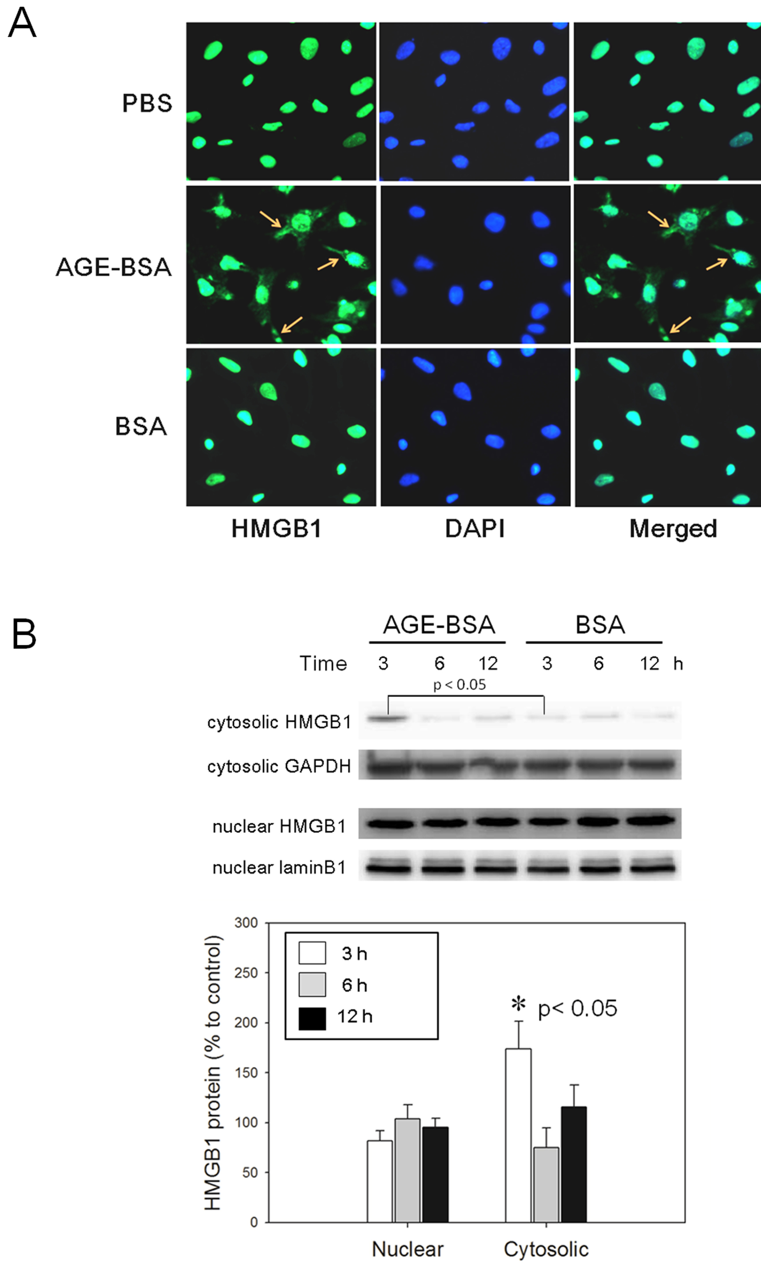
 Figure 2
of Lee, Mol Vis 2012; 18:838-850.
Figure 2
of Lee, Mol Vis 2012; 18:838-850.