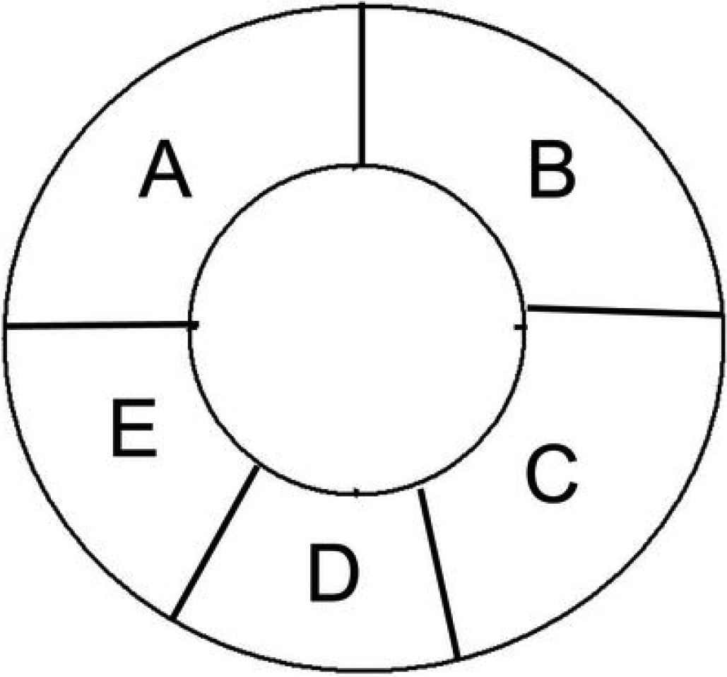
Figure 1. The corneal rims were
divided into five parts (A to E). Part A was used for
immunofluorescence staining. Part B was stained with trypan blue
and alizarin red. The endothelium from part C was stripped away
intact and frozen at −80 °C for RNA analysis. Part D and
part E were used for the immunohistochemical staining of p16INK4a
and Bmi1, respectively.

 Figure 1
of Wang, Mol Vis 2012; 18:803-815.
  Figure 1
of Wang, Mol Vis 2012; 18:803-815.