Figure 3. Nerve growth factor
promotes cell cycle progression via Akt and Erk activation in
human corneal epithelial cells. A: Human corneal
epithelial cells (HCECs) were incubated in keratinocyte
serum-free medium (K-SFM) without growth factors for 24 h before
treatment with nerve growth factor (NGF) at 25 ng/ml for 1 h. A
total of 50 µg cell lysates and total RNA were analyzed for
expression of the indicated genes by western blot analysis and
reverse transcriptase–polymerase chain reaction (RT–PCR),
respectively. Glyceraldehyde 3-phosphate dehydrogenase (GAPDH)
was used as a loading control. B: HCECs were incubated
in K-SFM without growth factors for 24 h and then pretreated
with LY294002 or PD98059 at 10 µM for 1 h before treatment with
NGF at 25 ng/ml for another hour. A total of 50 μg cell lysates
were analyzed for expression of the indicated genes by
immunoblotting analysis. GAPDH was used as a loading control.
Experiments were performed in triplicate. C: The
schematic representation depicts how the Akt and Erk pathways
collaborate to control NGF induction of cyclin D expression.
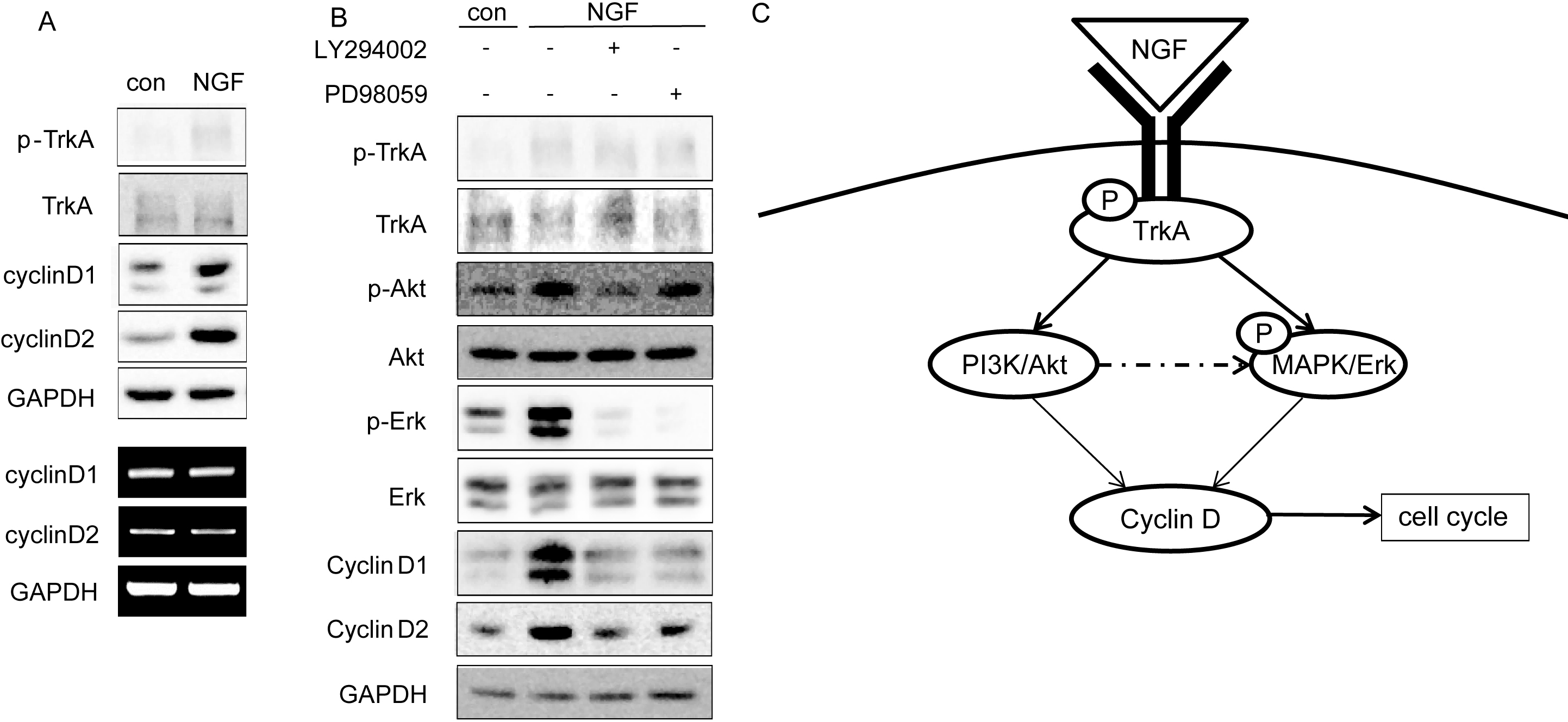
 Figure 3
of Hong, Mol Vis 2012; 18:758-764.
Figure 3
of Hong, Mol Vis 2012; 18:758-764.