Figure 1. Effect of nerve growth
factor on cell proliferation and the cell cycle in human corneal
epithelial cells. A: Human corneal epithelial cells
(HCECs) cultured in vitro displayed a polygonal pattern (left
panel). Immunostaining for cytokeratin 12 (green) followed by
Hoechst staining (blue; right panel) is shown. B: HCECs
were seeded onto 96 well plates at a density of 6×10³
per well in defined keratinocyte serum-free medium (K-SFM), and
then treated with recombinant nerve growth factor β-NGF at 5
ng/ml and subjected to a cell proliferation assay for up to 6
days. C: HCECs at passage 1 were plated onto 60-mm
dishes. The cell cycle was analyzed by flow cytometry with cells
treated identically at day 4 of the cell proliferation assay
(upper panel), and the percentage of each phase (G1-M)
is indicated on the right (lower panel). Experiments were
performed in triplicate and statistically analyzed with the
Student t test. The results are shown as mean±standard
deviation (SD). All p-values (*) were considered statistically
significant when p<0.05.
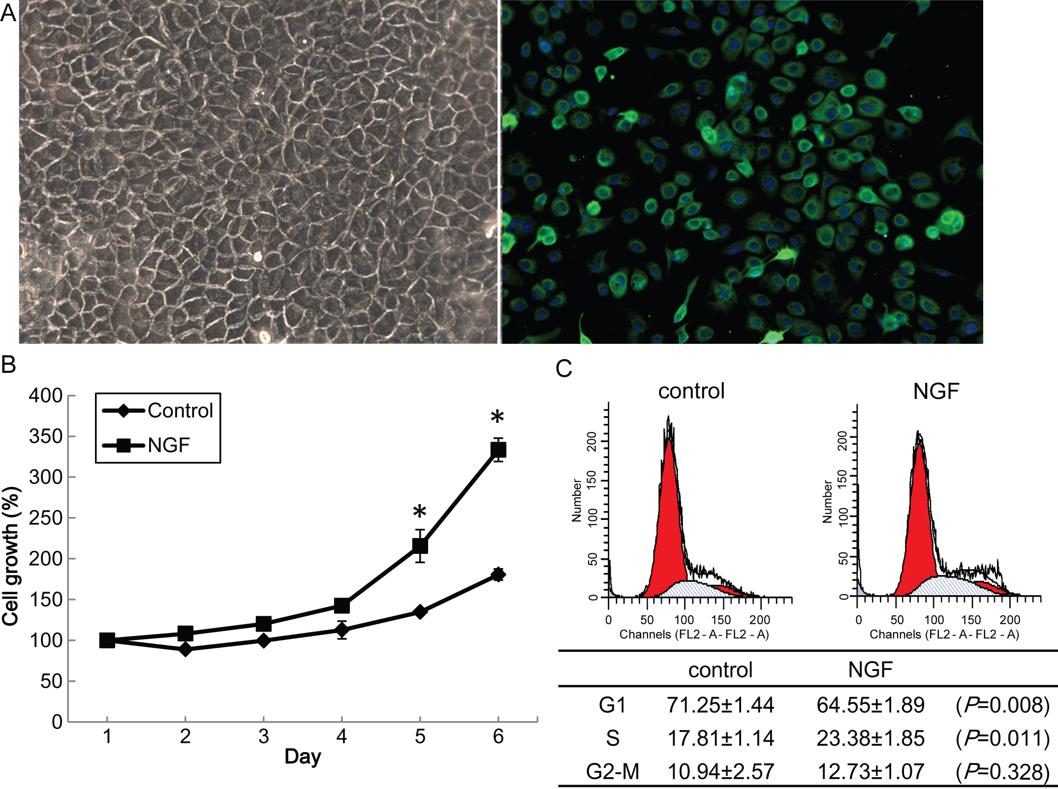
 Figure 1
of Hong, Mol Vis 2012; 18:758-764.
Figure 1
of Hong, Mol Vis 2012; 18:758-764.