Figure 1. Quantification of West Nile virus infection in ARPE-19 and human retinal pigment epithelial (hRPE) cells using flow cytometry.
Cells were infected with West Nile virus (WNV) at a multiplicity of infection (MOI) of 0.1, 1, and 10 (n=4), and after 24
h post infection, were fixed and permeabilized with the Cytofix/Cytoperm Fixation/Permeabilization Solution Kit (BD Biosciences)
and stained with fluorescein isothiocyanate (FITC)-conjugated antibodies specific for WNV nonstructural protein 1. Cells were
analyzed on a BD FACSCalibur flow cytometer. Data were analyzed using FlowJo (TreeStar software).
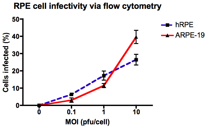
 Figure 1 of
Munoz-Erazo, Mol Vis 2012; 18:730-743.
Figure 1 of
Munoz-Erazo, Mol Vis 2012; 18:730-743.