Figure 1. Localization of SH3PXD2B in human eyes. Immunohistochemistry labeling of SH3PXD2B on human eyes reveals localization of SH3PXD2B in most ocular cell
types. (A-D, I-L) Cryosections were labeled with an anti-SH3PXD2B antibody (Green) and To-Pro-3, a nuclear counterstain (blue). (E-H, M-P) Negative controls omitting the primary antibody were performed on adjacent sections. (A-B, E-F) Cornea. (C, G) Iris. (D, H) Trabecular meshwork. (I, M) Ciliary processes. (J, N) Ciliary muscles. (K, O) Retina. (L, P) Lens. The orange-yellow color in K and O represents lipofuscin autofluorescence in the retinal pigment epithelium. cep, corneal epithelium; cen, corneal endothelium;
ist, iris stroma; ipe, iris pigment epithelium; tm, trabecular meshwork; sc, Schlemm’s canal; gcl, ganglion cell layer; inl,
inner nuclear layer; onl, outer nuclear layer; is, inner segment; os, outer segment. Scale bar=50 µm.
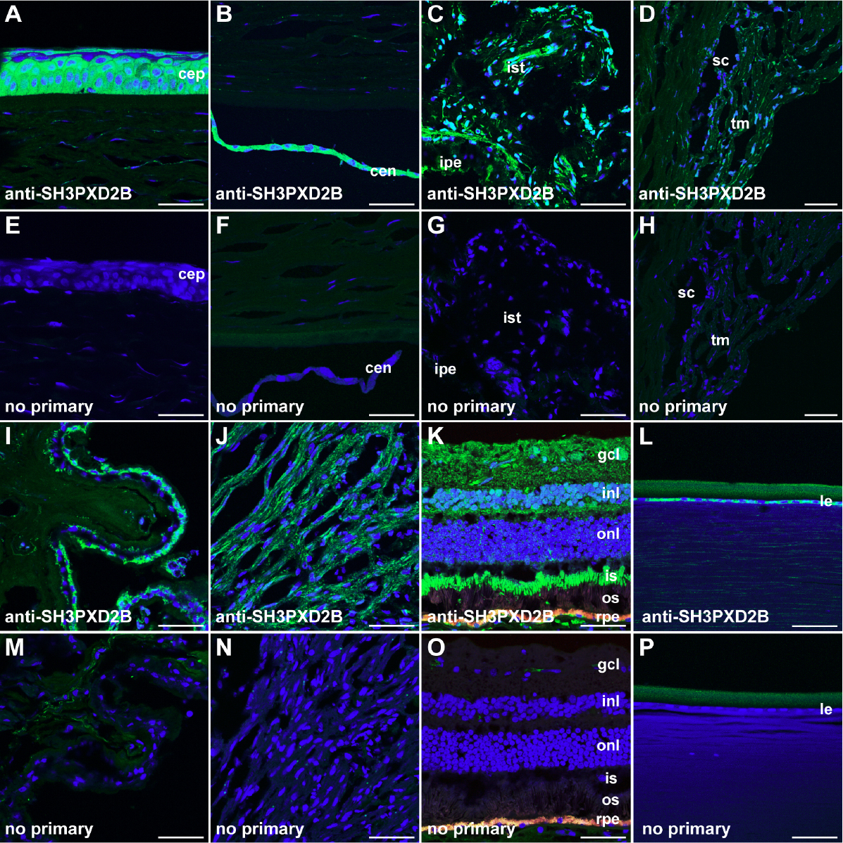
 Figure 1 of
Mao, Mol Vis 2012; 18:705-713.
Figure 1 of
Mao, Mol Vis 2012; 18:705-713.