Figure 3. Light damage is more severe
in the dorsal retina. Hematoxylin and eosin–stained sagittal
cross section of a left eye processed 14 days ALE. The superior
rectus muscle (black arrow) and the optic nerve (gray arrow) are
observed. The ellipse shows the region most affected by
phototoxicity, situated in the dorsal retina at approximately
400 microns from the optic disc. The insert shows a higher
magnification of this region, in which the thickness of the
outer segments of the photoreceptor layer (white arrow) and the
outer nuclear layer (ONL; asterisk) are very much reduced. D:
dorsal, V: ventral.
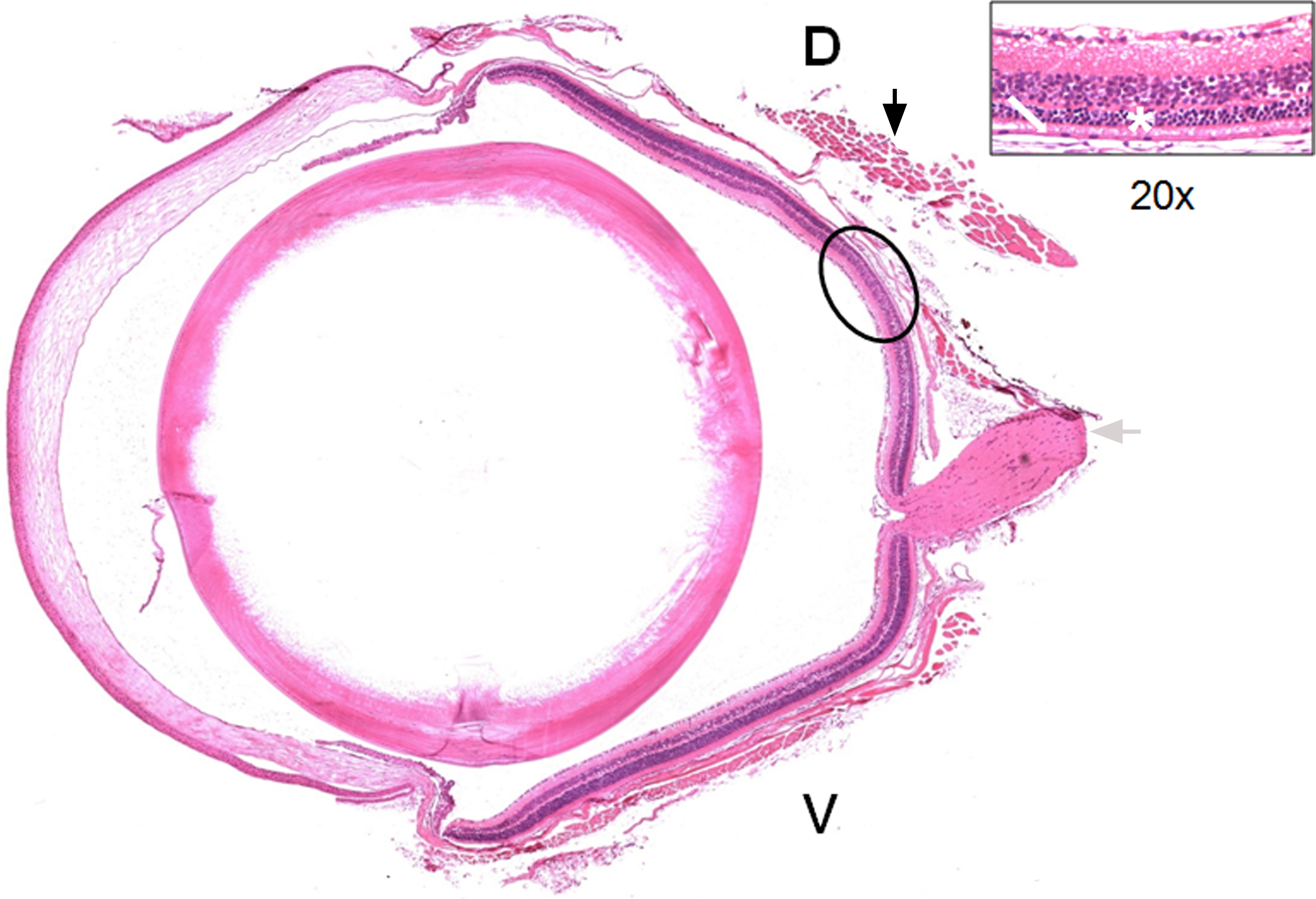
 Figure 3
of Montalbán-Soler, Mol Vis 2012;
18:675-693.
Figure 3
of Montalbán-Soler, Mol Vis 2012;
18:675-693.