Figure 2. Time course of retinal
degeneration after light exposure (ALE). Haematoxylin/eosin
stained retinal cross sections from the dorsal (left column) and
the ventral (right column) central retina. A, B:
control animals; C-L: experimental retinas from
animals processed at increasing times ALE. C, D:
1 day ALE; E, F: 7 days ALE; G, H:
30 days ALE; I, J: 90 days ALE and K, L:
180 days ALE. Abbreviations: RGCL: retinal ganglion cell layer,
IPL: inner plexiform layer, INL: inner nuclear layer, OPL: outer
plexiform layer, ONL: outer nuclear layer, OSPL: outer segment
of photoreceptors layer. Asterisks mark the outer nuclear layer
and the arrows point to the outer segments of photoreceptors.
Bar=100 μm.
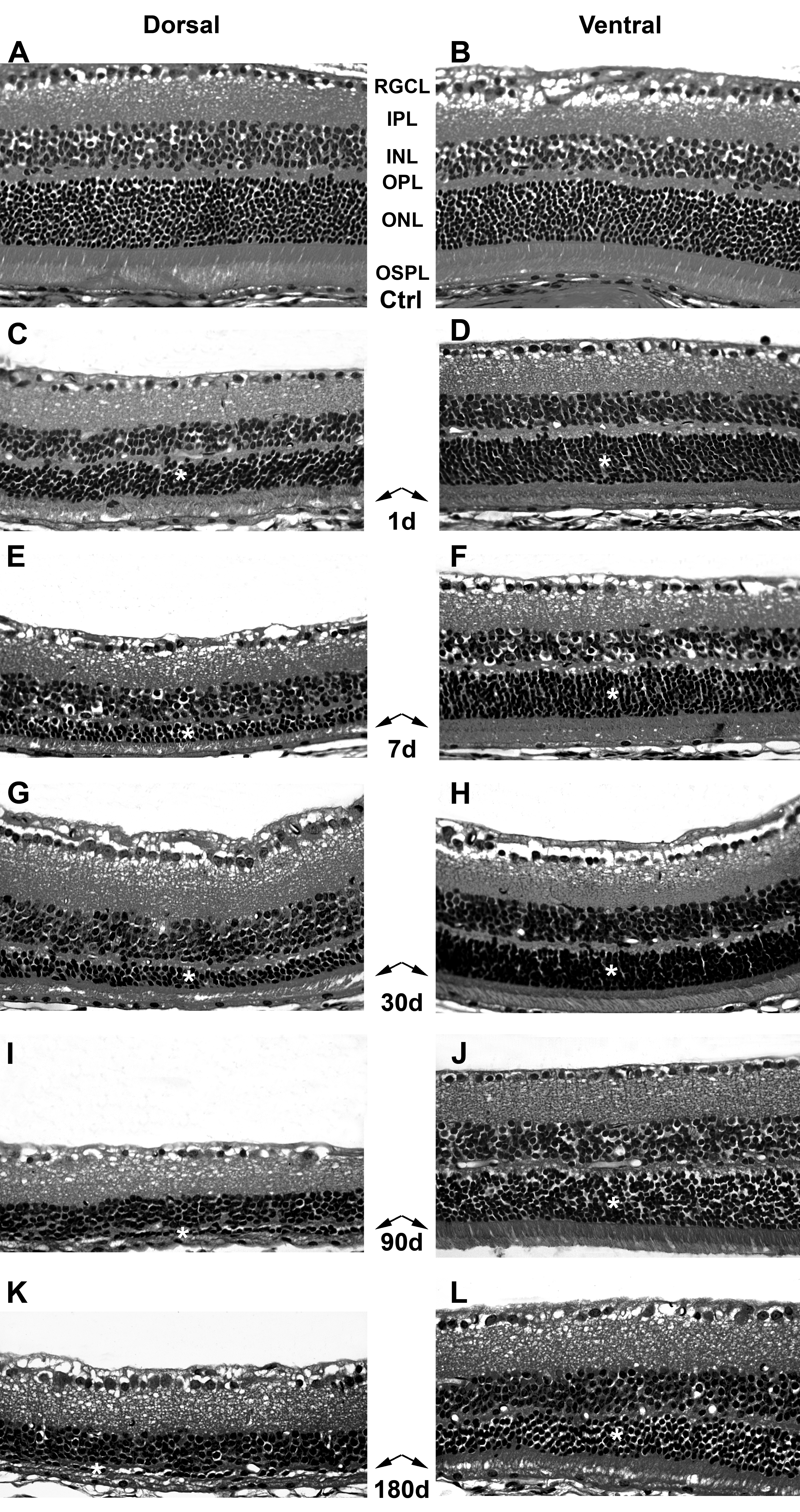
 Figure 2
of Montalbán-Soler, Mol Vis 2012;
18:675-693.
Figure 2
of Montalbán-Soler, Mol Vis 2012;
18:675-693.